Review
. 2017 Jan;37(1):26-34.
doi: 10.1161/ATVBAHA.116.303229. Epub 2016 Nov 22.

Altered Smooth Muscle Cell Force Generation as a Driver of Thoracic Aortic Aneurysms and Dissections

Affiliations
Review

Altered Smooth Muscle Cell Force Generation as a Driver of Thoracic Aortic Aneurysms and Dissections

Dianna M Milewicz et al. Arterioscler Thromb Vasc Biol. 2017 Jan.

Erratum in

  • Correction.
    [No authors listed] [No authors listed] Arterioscler Thromb Vasc Biol. 2017 Feb;37(2):e12. doi: 10.1161/ATV.0000000000000045. Arterioscler Thromb Vasc Biol. 2017. PMID: 28122778 No abstract available.

Abstract

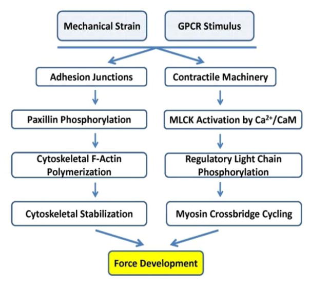
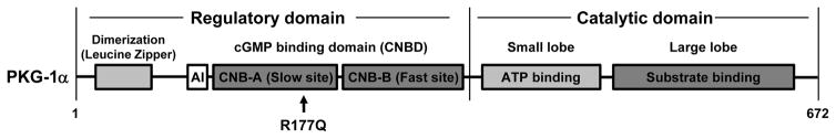
The importance of maintaining contractile function in aortic smooth muscle cells (SMCs) is evident by the fact that heterozygous mutations in the major structural proteins or kinases controlling contraction lead to the formation of aneurysms of the ascending thoracic aorta that predispose to life-threatening aortic dissections. Force generation by SMC requires ATP-dependent cyclic interactions between filaments composed of SMC-specific isoforms of α-actin (encoded by ACTA2) and myosin heavy chain (MYH11). ACTA2 and MYH11 mutations are predicted or have been shown to disrupt this cyclic interaction predispose to thoracic aortic disease. Movement of the myosin motor domain is controlled by phosphorylation of the regulatory light chain on the myosin filament, and loss-of-function mutations in the dedicated kinase for this phosphorylation, myosin light chain kinase (MYLK) also predispose to thoracic aortic disease. Finally, a mutation in the cGMP-activated protein kinase (PRKG1) results in constitutive activation of the kinase in the absence of cGMP, thus driving SMC relaxation in part through increased dephosphorylation of the regulatory light chain and predisposes to thoracic aortic disease. Furthermore, SMCs cannot generate force without connections to the extracellular matrix through focal adhesions, and mutations in the major protein in the extracellular matrix, fibrillin-1, linking SMCs to the matrix also cause thoracic aortic disease in individuals with Marfan syndrome. Thus, disruption of the ability of the aortic SMC to generate force through the elastin-contractile units in response to pulsatile blood flow may be a primary driver for thoracic aortic aneurysms and dissections.

Keywords: Marfan syndrome; aortic aneurysm; aortic dissection; mutation; thoracic aorta; vascular smooth muscle cells.


Figures

Figure 1
Figure 1
Signaling Pathways Controlling Regulatory Light Chain (RLC) Phosphorylation and Activation of Vascular Smooth Muscle Contraction and Relaxation. Contraction pathways shown on the left lead to calcium-dependent phosphorylation of myosin RLC by myosin light chain kinase (MLCK) and actin-myosin-dependent force development. Relaxation pathways on the right lead to calcium reduction and myosin dephosphorylation. Mutated genes causing heritable thoracic aortic disease are indicated in red font; these genes disrupt critical proteins involved in SMC contraction. Abbreviations: BK (large-conductance calcium and voltage sensitive potassium channel), CaM (calmodulin), GP (Guanine nucleotide-binding Protein, e.g. Gq/11), GPCR (GP-Coupled Receptor, e.g. α1 adrenergic receptor), IP3R (Inositol triphosphate Receptor), IRAG (Inositol triphosphate Receptor-Associated PKG substrate), LIC (Ligand-gated Ion Channel, e.g. ATP-gated P2X1), RLC (Regulatory Light Chain), -P (phosphorylation), MADP (Membrane-Associated Dense Plaque), PKs (Protein Kinases, e.g. Rho kinase), SR (sarcoplasmic reticulum), VDCC (Voltage-Dependent Calcium Channel, e.g. CaV1.2). Red lightning bolt represents membrane depolarization.
Figure 2
Figure 2
Vascular smooth muscle force development is dependent on submembranous cytoskeletal actin polymerization initiated at focal adhesions and actomyosin cross-bridge cycling driven by phosphorylation of the myosin regulatory light chain.
Figure 3
Figure 3
Missense mutations in ACTA2 identified in individuals and families with heritable thoracic aortic disease. Residues highlighted in boxes have the following correlations between genotype and phenotype: R179 mutations cause Multisystemic Smooth Muscle Dysfunction Syndrome; R258 and R39 mutations predispose to a moyamoya-like cerebrovascular disease; R149 and R118 predispose to early onset coronary artery disease; and G160 and W88 disruption predispose to decreased penetrance and later onset thoracic aortic disease. Side chains of other residues known to be mutated are shown as sticks (cyan). G48V and M49V are in a disordered region (D-loop) and not seen in this crystal structure. Actin structure is PDB code 1NWK.
Figure 4
Figure 4
Schematic of a SMC myosin filament with the location of the regulatory light chain (RLC) and essential light chain (ELC) and protein domains of the myosin heavy chain indicated. In frame deletions in the coiled coil domain predispose to heritable thoracic aortic disease.
Figure 5
Figure 5
Schematic of the protein domains in the three isoforms of myosin light chain kinase (MLCK). Three distinct promoters control the expression of these isoforms of MLCK. The short form is expression in the aorta. Pathogenic mutations that cause heritable thoracic aortic disease are shown and lead to haploinsufficiency or disrupt calmodulin binding, which is required to activate the kinase.
Figure 6
Figure 6
Schematic of type I cGMP-dependent protein kinase (PKG-1), which is activated upon binding of cGMP to the CNB-A and CNB-B domains. With cGMP binding, the autoinhibitory domain (AI) is released from the catalytic domain to activate the kinase. A single mutation, R177Q, has been identified to cause heritable thoracic aortic disease and is located in the CNB-A cGMP binding domain. The mutation leads to constitutive activation of the kinase such that it is no longer regulated by cGMP.

References

    1. LeMaire SA, Russell L. Epidemiology of thoracic aortic dissection. Nat Rev Cardiol. 2011 Feb;8:103–13. - PubMed
    1. Howard DP, Banerjee A, Fairhead JF, Perkins J, Silver LE, Rothwell PM. Population-based study of incidence and outcome of acute aortic dissection and premorbid risk factor control: 10-year results from the Oxford Vascular Study. Circulation. 2013 May 21;127:2031–7. - PMC - PubMed
    1. Sakai LY, Keene DR, Renard M, De BJ. FBN1: The disease-causing gene for Marfan syndrome and other genetic disorders. Gene. 2016 Oct 10;591:279–91. - PMC - PubMed
    1. Guo DC, Pannu H, Papke CL, et al. Mutations in smooth muscle alpha-actin (ACTA2) lead to thoracic aortic aneurysms and dissections. Nat Genet. 2007 Dec;39:1488–93. - PubMed
    1. Zhu L, Vranckx R, Khau Van KP, Lalande A, Boisset N, Mathieu F, Wegman M, Glancy L, Gasc JM, Brunotte F, Bruneval P, Wolf JE, Michel JB, Jeunemaitre X. Mutations in myosin heavy chain 11 cause a syndrome associating thoracic aortic aneurysm/aortic dissection and patent ductus arteriosus. Nat Genet. 2006 Mar;38:343–9. - PubMed

Publication types

MeSH terms