Smooth muscle tumour of uncertain malignant potential (SMTUMP) in the nasal cavity: an incidental finding
- PMID: 27879303
- PMCID: PMC5129059
- DOI: 10.1136/bcr-2016-217386
Smooth muscle tumour of uncertain malignant potential (SMTUMP) in the nasal cavity: an incidental finding
Abstract
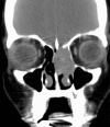
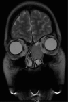
Sino-nasal smooth muscle tumours of uncertain malignant potential (SMTUMP) are very rare neoplasms of mesenchymal origin with features in between a benign leiomyoma and a leiomyosarcoma. We report a rare case of SMTUMP in a 44-year-old woman, who presented with vague symptoms of pharyngitis. Nasal endoscopy revealed a smooth mass in left nasal cavity. Contrast-enhanced CT and MRI scans showed features likely to be inverted papilloma or olfactory neuroblastoma or meningioma. Excision was planned and intraoperatively, frozen section revealed a probable spindle cell lesion. Final histopathological report following immunohistochemistry (IHC) & immunofluoresence (IF) confirmed it to be a SMTUMP. This patient underwent complete resection via endoscopic KTP laser assisted, anterior skull base surgery with no recurrence on follow-up.
2016 BMJ Publishing Group Ltd.
Conflict of interest statement
Conflicts of Interest: None declared.
Figures


References
-
- Huang CT, Chien CY, Su CY et al. . Leiomyoma of the inferior turbinates. J Otolaryngol 2000;29:55–6. - PubMed
-
- Huang HY, Antonescu CR. Sinonasal smooth muscle cell tumors: a clinicopathologic and immunohistochemical analysis of 12 cases with emphasis on the low-grade end of the spectrum. Arch Pathol Lab Med 2003;127:297–304. doi:10.1043/0003-9985(2003)127<0297:SSMCT>2.0.CO;2 - DOI - PubMed
-
- Kuruvilla A. Leiomyosarcoma of the sinonasal tract: a clinicopathological study of nine cases. Arch Otolaryngol Head Neck Surg 1990;27:129–35. - PubMed
-
- Nicolai P, Redaelli de Zinis LO, Facchetti F et al. . Craniofacial resection for vascular leiomyoma of the nasal cavity. Am J Otolaryngol 1996;17:340–4. - PubMed
-
- Trott MS, Gewirtz A, Lavertu P et al. . Sinonasal leiomyomas. Otolaryngol Head Neck Surg 1994;111:660–4. - PubMed
Publication types
MeSH terms
LinkOut - more resources
Full Text Sources
Other Literature Sources
Medical
Miscellaneous
