. 2017 Jan 6;292(1):292-304.
doi: 10.1074/jbc.M116.763268. Epub 2016 Nov 22.

AMP-activated Kinase (AMPK) Promotes Innate Immunity and Antiviral Defense through Modulation of Stimulator of Interferon Genes (STING) Signaling

Affiliations

AMP-activated Kinase (AMPK) Promotes Innate Immunity and Antiviral Defense through Modulation of Stimulator of Interferon Genes (STING) Signaling

Daniel Prantner et al. J Biol Chem. .

Abstract

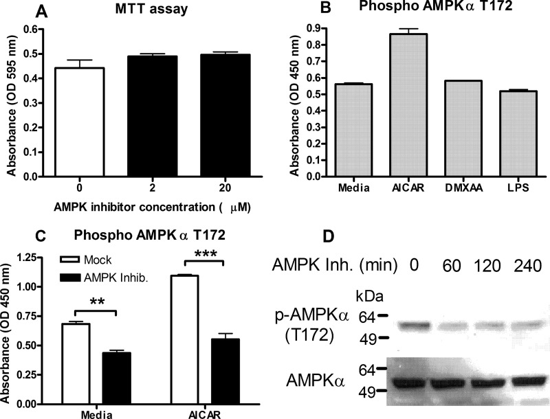
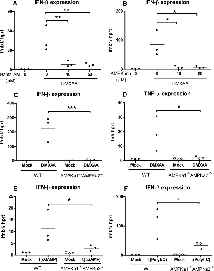
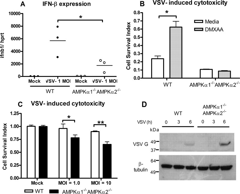
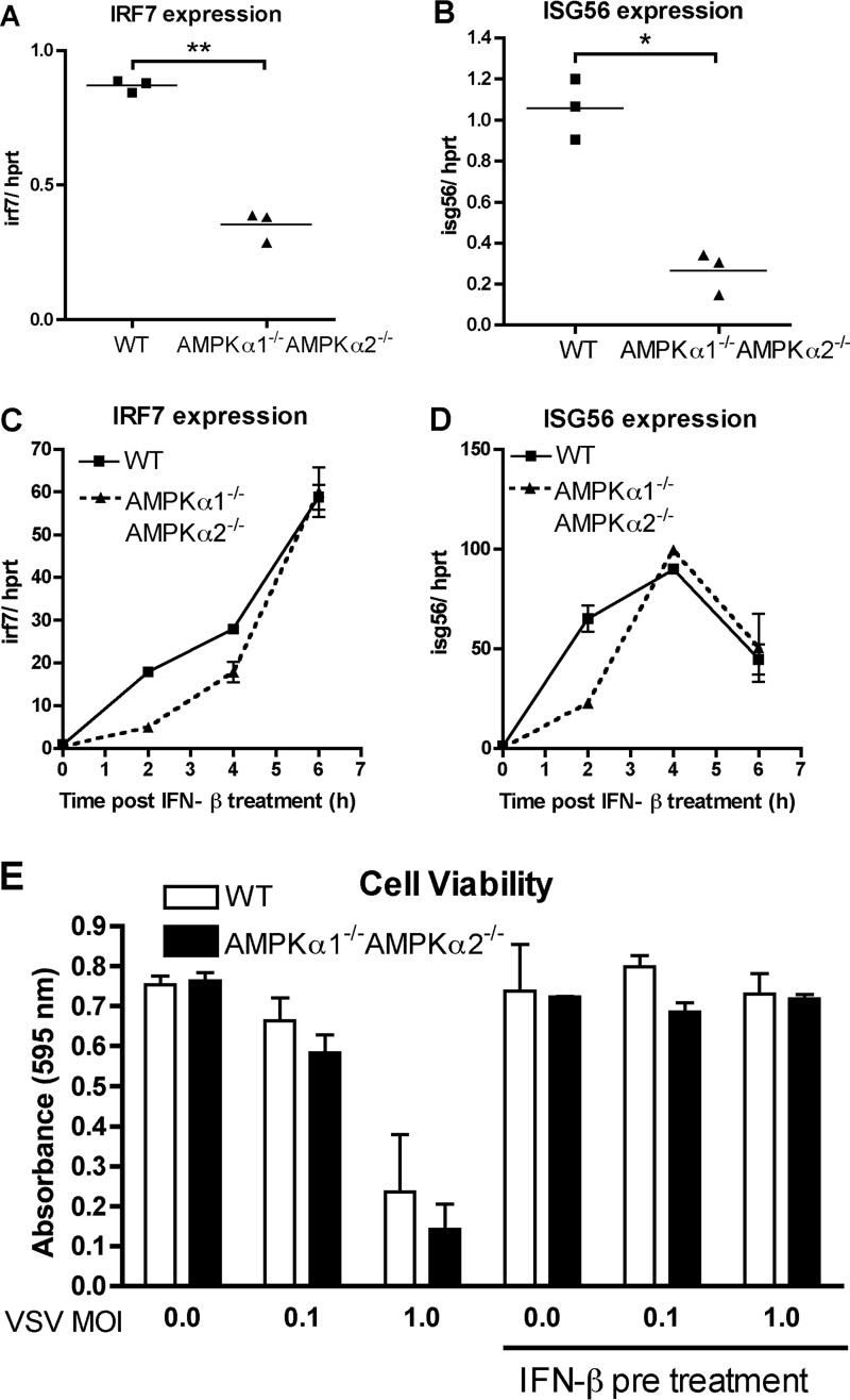
The host protein Stimulator of Interferon Genes (STING) has been shown to be essential for recognition of both viral and intracellular bacterial pathogens, but its regulation remains unclear. Previously, we reported that mitochondrial membrane potential regulates STING-dependent IFN-β induction independently of ATP synthesis. Because mitochondrial membrane potential controls calcium homeostasis, and AMP-activated protein kinase (AMPK) is regulated, in part, by intracellular calcium, we postulated that AMPK participates in STING activation; however, its role has yet to be been defined. Addition of an intracellular calcium chelator or an AMPK inhibitor to either mouse macrophages or mouse embryonic fibroblasts (MEFs) suppressed IFN-β and TNF-α induction following stimulation with the STING-dependent ligand 5,6-dimethyl xanthnone-4-acetic acid (DMXAA). These pharmacological findings were corroborated by showing that MEFs lacking AMPK activity also failed to up-regulate IFN-β and TNF-α after treatment with DMXAA or the natural STING ligand cyclic GMP-AMP (cGAMP). As a result, AMPK-deficient MEFs exhibit impaired control of vesicular stomatitis virus (VSV), a virus sensed by STING that can cause an influenza-like illness in humans. This impairment could be overcome by pretreatment of AMPK-deficient MEFs with type I IFN, illustrating that de novo production of IFN-β in response to VSV plays a key role in antiviral defense during infection. Loss of AMPK also led to dephosphorylation at Ser-555 of the known STING regulator, UNC-51-like kinase 1 (ULK1). However, ULK1-deficient cells responded normally to DMXAA, indicating that AMPK promotes STING-dependent signaling independent of ULK1 in mouse cells.

Keywords: AMP-activated kinase (AMPK); cytokine induction; interferon; macrophage; signal transduction.


Figures

FIGURE 1.
FIGURE 1.
Intracellular calcium concentration regulates STING-dependent cytokine induction. Peritoneal mouse macrophages were pretreated with the indicated dose of the intracellular calcium chelator BAPTA-AM or vehicle alone for 30 min. The cells were then treated for an additional 2 h with DMXAA (100 μg/ml). Expression of IFN-β (A) and TNF-α (B) mRNA were determined by qRT-PCR. The data represents the mean fold-induction calculated from 3 independent experiments. The data points represent the individual mean from each experiment. **, p < 0.01; ***, p < 0.001.
FIGURE 2.
FIGURE 2.
Inhibition of AMPK activity blocks induction of STING-dependent cytokines. Peritoneal macrophages were treated with the indicated doses of the AMPK inhibitor Compound C or vehicle alone for 30 min. The cells were then treated for an additional 2 h with DMXAA (100 μg/ml). Expression of IFN-β (A) and TNF-α (B) mRNA were determined by qRT-PCR. The data represents the mean fold-induction calculated from 3 independent experiments. The data points represent the individual mean from each experiment. NS, not significantly different. *, p < 0.05; **, p < 0.01; ***, p < 0.001.
FIGURE 3.
FIGURE 3.
AMPK inhibition lowers basal and inducible activities of the AMPK signaling complex. Peritoneal macrophages were treated with the indicated doses of the AMPK inhibitor Compound C for 2 h. At the end of this incubation, mitochondrial dehydrogenase activity (A) was quantified by using the MTT assay described under “Experimental Procedures.” B, peritoneal macrophages were treated with AICAR (0.5 mm), DMXAA (100 μg/ml), LPS (100 ng/ml), or media only for 30 min. C, peritoneal macrophages were pre-treated with Compound C (20 μm) for 30 min prior to stimulation with AICAR (0.5 mm) or solvent alone for an additional 30 min. Phosphorylation of the Thr-172 amino acid residue of AMPKα was determined by an ELISA-based assay as described under “Experimental Procedures” in B and C. The data represents the mean ± S.D. for experimental conditions performed in triplicate (A) or duplicate (B and C). A representative of 3 independent experiments is shown. **, p < 0.01; ***, p < 0.001. D, peritoneal macrophages were stimulated for the indicated times with Compound C (20 μm). Lysates were separated by SDS-PAGE. After transfer, proteins were detected by Western blot analysis using antibodies specific for phospho-AMPK (Thr-172) or total AMPK.
FIGURE 4.
FIGURE 4.
Cells deficient for AMPK signaling exhibit impaired cytokine induction mediated through cytosolic pathogen recognition receptors. A, WT MEFs were treated with the indicated doses of the intracellular calcium chelator BAPTA-AM or solvent alone for 30 min. The cells were then treated for an additional 2 h with DMXAA (100 μg/ml). B, cells were treated as in A except with the AMPK inhibitor Compound C or solvent alone prior to DMXAA treatment. Expression of IFN-β (A and B) was determined by qRT-PCR. C and D, WT or AMPKα1−/−AMPKα2−/− MEFs were stimulated for 2 h with DMXAA (100 μg/ml). Expression of IFN-β (C) and TNF-α (D) mRNA were determined by qRT-PCR. E, WT and AMPKα1−/−AMPKα2−/− MEFs were transfected with 1 μg of cGAMP or 1 μg of poly(I:C) and incubated for 6 h. Expression of IFN-β mRNA (E and F) was determined by qRT-PCR. For A–E, the data represents the mean fold-induction from 2 or 3 independent experiments. *, p < 0.05; **, p < 0.01; ***, p < 0.001.
FIGURE 5.
FIGURE 5.
Cells deficient for AMPK signaling exhibit impaired STING-dependent signaling. WT or AMPKα1−/−AMPKα2−/− MEFs were stimulated for the indicated times with DMXAA (100 μg/ml). Lysates were separated by SDS-PAGE. After transfer, proteins were detected by Western blot analysis using antibodies specific for phospho-TBK1, total TBK1, phospho-IRF3, IRF3, and β-Tubulin. A representative of 3 independent experiments is shown.
FIGURE 6.
FIGURE 6.
Decreased IFN-β response to VSV infection increases susceptibility of AMPK-deficient cells. WT and AMPKα1−/−AMPKα2−/− MEFs were infected with VSV (m.o.i. = 1) for 6 h. Prior to onset of cell death at 6 h post-infection, IFN-β mRNA (A) expression was quantified by qRT-PCR. For A, the data represents the mean fold-induction from 3 independent experiments. B, WT and AMPKα1−/−AMPKα2−/− MEFs were pretreated with DMXAA (100 μg/ml) or vehicle only for 6 h. Immediately thereafter, cells were infected with VSV (m.o.i. = 10) for 18 h. Viability was determined using the MTT assay and normalized to mock-infected cells to give a cell survival index. C, WT and AMPKα1−/−AMPKα2−/− MEFs were infected with the indicated m.o.i. of VSV. At 8 h post-infection, viability was calculated as in B. For B and C, the data represents the mean ± S.D. for experimental conditions performed in duplicate in the same experiment. *, p < 0.05; **, p < 0.01. D, WT MEFs and AMPKα1−/−AMPKα2−/− MEFs were infected with VSV (m.o.i. = 1). At the indicated time points, cell lysates were harvested and separated by SDS-PAGE. After transfer, proteins were detected by Western blot analysis using antibodies specific for VSV G protein or β-tubulin. A representative of 3 independent experiments is shown.
FIGURE 7.
FIGURE 7.
Cells deficient for AMPK signaling exhibit normal responses to exogenous type I IFN. Expression of the ISGs IRF7 (A) and ISG56 (B) mRNA in untreated WT MEFs or AMPKα1−/−AMPKα2−/− MEFs were examined. Each data point represents the pooled results from 3 independent experiments. Induction of the ISGs IRF7 (C) and ISG56 (D) in WT or AMPKα1−/−AMPKα2−/− MEFs were monitored for 6 h following stimulation with IFN-β (100 units/ml). E, WT and AMPKα1−/−AMPKα2−/− MEFs were pretreated with IFN-β (100 units/ml) or vehicle only for 6 h. Immediately thereafter, cells were infected with VSV at the indicated m.o.i. for 18 h. Viability was calculated using the MTT assay. The data represents the mean ± S.D. for experimental conditions performed in triplicate for panels C–E and a representative of 3 independent experiments is shown. *, p = 0.0016; **, p = 0.0001.
FIGURE 8.
FIGURE 8.
Cells deficient for AMPK signaling have constitutively dephosphorylated ULK1. WT and AMPKα1−/−AMPKα2−/− MEFs were stimulated for the indicated times with DMXAA (100 μg/ml). Lysates were separated by SDS-PAGE. After transfer, proteins were detected by Western blot analysis using antibodies specific for phospho-ULK1 (Ser-555), total ULK1, and β-Tubulin. A representative of 3 independent experiments is shown.
FIGURE 9.
FIGURE 9.
The ULK1 inhibitor SBI-0206965 represses activation of STING-dependent signaling. WT MEFs were treated for 30 min with the ULK1 inhibitor SBI-0206965 (50 μm) or vehicle alone prior to treatment with DMXAA (100 μg/ml) for 2 h. Expression of IFN-β mRNA (A) was determined by qRT-PCR. The data in A represents the mean fold-induction calculated from 3 independent experiments. The data points represent the individual means from each experiment. B, WT MEFs were treated with the ULK1 inhibitor SBI-0206965 (50 μm) or vehicle alone or 2 h. At the end of this incubation, mitochondrial dehydrogenase activity was quantified by using the MTT assay as described under “Experimental Procedures.” The data in B represents the mean ± S.D. for experimental conditions performed in triplicate.
FIGURE 10.
FIGURE 10.
Absence of ULK1 does not impair STING-dependent signaling. A, WT and ULK1−/− MEFs were genotyped by probing cellular lysates with antibodies specific for ULK1 and β-tubulin. B, WT and ULK1−/− MEFs were stimulated by transfecting 1 μg of cGAMP into the cells and incubated for 6 h. C, WT and ULK1−/− MEFs were stimulated for 2 h with DMXAA (100 μg/ml). D, cells were treated as in C, but were also pretreated for 30 min with the indicated dose of SBI-0206965. In B–D, expression of IFN-β was determined by qRT-PCR. The data in B–D, represents the mean fold-induction calculated from 3 independent experiments.
FIGURE 11.
FIGURE 11.
Multiple regulatory pathways are utilized by the cell to control activation of the STING signaling pathway. A, our data support a model in which the presence of AMPK signaling is required for optimal activation of STING-dependent signaling. Conditions that interfere with AMPK signaling, including chelation of intracellular calcium (1), pharmacologic inhibition of AMPK signaling (2), or genetic ablation of AMPK signaling (3) inhibit STING-dependent signaling. As ULK1−/− and ULK1−/−ULK2−/− MEFs exhibit no defect in response to STING agonists, we conclude that the regulatory role of AMPK on STING is independent of ULK1 and that the ULK1 inhibitor SBI-0206965 has off-target effects leading to signal impairment. B, the exact role of AMPK in the regulation of STING is unknown at present. AMPK might directly promote STING signaling by substrate level phosphorylation (1). Alternatively, it might act indirectly by activating a positive regulator (PR) or inhibiting a negative regulator (NR) of STING-dependent signaling (2). An alternate possibility is that the STING signaling pathway operates efficiently in a narrow metabolic window and is indirectly inhibited in the absence of the catabolic pathways promoted by AMPK (3). Future studies will delineate which of these mechanisms is operative.

Similar articles

Cited by

References

    1. Ishikawa H., and Barber G. N. (2008) STING is an endoplasmic reticulum adaptor that facilitates innate immune signalling. Nature 455, 674–678 - PMC - PubMed
    1. Zhong B., Yang Y., Li S., Wang Y. Y., Li Y., Diao F., Lei C., He X., Zhang L., Tien P., and Shu H. B. (2008) The adaptor protein MITA links virus-sensing receptors to IRF3 transcription factor activation. Immunity 29, 538–550 - PubMed
    1. Sun W., Li Y., Chen L., Chen H., You F., Zhou X., Zhou Y., Zhai Z., Chen D., and Jiang Z. (2009) ERIS, an endoplasmic reticulum IFN stimulator, activates innate immune signaling through dimerization. Proc. Natl. Acad. Sci. U.S.A. 106, 8653–8658 - PMC - PubMed
    1. Gao D., Wu J., Wu Y. T., Du F., Aroh C., Yan N., Sun L., and Chen Z. J. (2013) Cyclic GMP-AMP synthase is an innate immune sensor of HIV and other retroviruses. Science 341, 903–906 - PMC - PubMed
    1. Ishikawa H., Ma Z., and Barber G. N. (2009) STING regulates intracellular DNA-mediated, type I interferon-dependent innate immunity. Nature 461, 788–792 - PMC - PubMed

MeSH terms