. 2017 Jan;173(1):582-599.
doi: 10.1104/pp.16.01602. Epub 2016 Nov 22.

Phosphorylation of MAP65-1 by Arabidopsis Aurora Kinases Is Required for Efficient Cell Cycle Progression

Affiliations

Phosphorylation of MAP65-1 by Arabidopsis Aurora Kinases Is Required for Efficient Cell Cycle Progression

Joanna Boruc et al. Plant Physiol. 2017 Jan.

Abstract

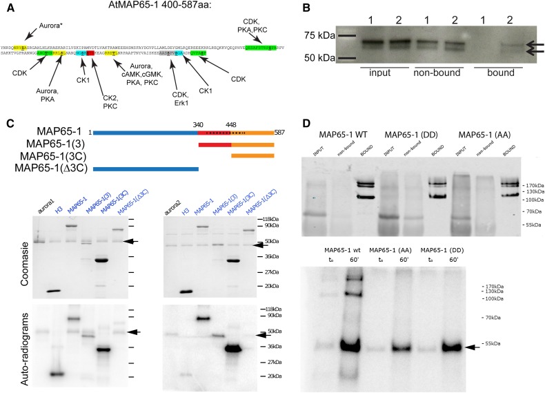
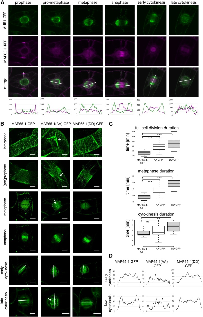
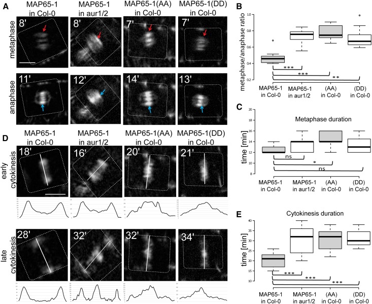
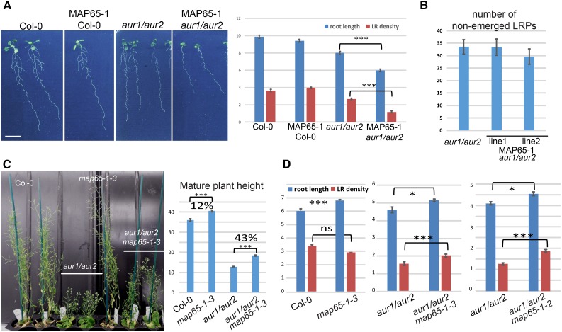
Aurora kinases are key effectors of mitosis. Plant Auroras are functionally divided into two clades. The alpha Auroras (Aurora1 and Aurora2) associate with the spindle and the cell plate and are implicated in controlling formative divisions throughout plant development. The beta Aurora (Aurora3) localizes to centromeres and likely functions in chromosome separation. In contrast to the wealth of data available on the role of Aurora in other kingdoms, knowledge on their function in plants is merely emerging. This is exemplified by the fact that only histone H3 and the plant homolog of TPX2 have been identified as Aurora substrates in plants. Here we provide biochemical, genetic, and cell biological evidence that the microtubule-bundling protein MAP65-1-a member of the MAP65/Ase1/PRC1 protein family, implicated in central spindle formation and cytokinesis in animals, yeasts, and plants-is a genuine substrate of alpha Aurora kinases. MAP65-1 interacts with Aurora1 in vivo and is phosphorylated on two residues at its unfolded tail domain. Its overexpression and down-regulation antagonistically affect the alpha Aurora double mutant phenotypes. Phospho-mutant analysis shows that Aurora contributes to the microtubule bundling capacity of MAP65-1 in concert with other mitotic kinases.


Figures

Figure 1.
Figure 1.
Arabidopsis MAP65-1 is a substrate of alpha Aurora kinases. A, The C-terminal domain of Arabidopsis MAP65-1 carries multiple putative phosphorylation sites (colored). Within each peptide sequence the phosphorylatable S/T residue is highlighted in bold and underlined. All putative kinase motifs (except for the first predicted Aurora site, marked with a star) were identified in Smertenko et al. (2006). The three putative Aurora phosphorylation sites (marked in yellow) were predicted based on the Aurora consensus phosphorylation motif identified in budding yeast Ipl1 (Cheeseman et al., 2002) and human Aurora A (Ferrari et al., 2005). B, Co-immunoprecipitation of MAP65-1 from control Col-0 plants (1) and complemented aur1/aur2 double knockdown mutant seedlings expressing genomic Aurora1-GFP (2). In contrast to the control, endogenous MAP65-1 (present as two bands and detected using a MAP65-1 antibody) is specifically pulled down with AUR1-GFP in the bound fraction (indicated with the arrows). C, Schematic representation of MAP65-1 [Projection domain (blue), C-terminal moiety (red and orange) with an unfolded divergent C terminus domain (orange), spectrin-like domain (dashed black bar)] and the truncations used for the in vitro phosphorylation assays using recombinant Arabidopsis Aurora1 (left) and Aurora2 (right). The alpha Auroras phosphorylate MAP65-1 at its C-terminal domain (domain 3C). Top gel pictures are Coomassie staining, bottom gel pictures are autoradiograms. Phosphorylation of histone H3 was included as a positive control for Aurora phosphorylation. Arrows mark the position of Aurora1 and Aurora2. D, GFP-fused MAP65-1 isoforms (wild type, AA, and DD), expressed in Nicotiana benthamiana leaves were immunoprecipitated and confirmed using Western blot (upper panel). The higher MW bands at 170 probably represent MAP65-1 dimers. In vitro Aurora1 kinase assays using the immunoprecipitated GFP-fused MAP65-1 isoforms as substrates (autoradiogram, lower panel, t0 and t60 min) show that wild-type MAP65-1 is phosphorylated by Aurora1, in contrast to the Aurora phosho-mimicking form [MAP65-1(DD) and the nonphosphorylatable form MAP65-1(AA)]. The arrow marks the autophosphorylation of Aurora1.
Figure 2.
Figure 2.
Phospho-mutations at S532 and T552 alter MAP65-1 properties in vivo. A, Colocalization between Aurora1-GFP and MAP65-1-RFP throughout mitosis. The panels are taken from three different time-lapse movies. The fluorescence intensity plots (shown below the merged images) demonstrate colocalization of Aurora1-GFP and MAP65-1-RFP e.g. at the nuclear envelope during the PPB stage and during cytokinesis as well as a negative correlation between Aurora1 localization and MAP65-1 MT bundling from prophase to cytokinesis. Scale bar = 10 μm. B, Representative images (n > 10) of Arabidopsis MAP65-1-GFP, MAP65-1(AA)-GFP, and MAP65-1(DD)-GFP expressed in tobacco BY-2 cells during consecutive stages of cell division. Arrows indicate excessive microtubule bundling observed in prophase, metaphase, and late cytokinesis in cells expressing MAP65-1(AA)-GFP. Images of cells in interphase and preprophase are projections, while the other phases are represented by single images. Scale bar = 10 μm. C, Box plot representation of full cell division (top), metaphase (middle), and cytokinesis (bottom) duration in BY-2 cells expressing MAP65-1-GFP (n = 13), MAP65-1(AA)-GFP (n = 14), and MAP65-1(DD)-GFP (n = 9). Overexpression of MAP65-1(AA), but also MAP65-1(DD) prolongs cell division duration compared to the wild-type MAP65-1 (t test; triple asterisk: P < 0.0001; double asterisk: P < 0.001; ns (not significant): P > 0.01). Center lines show the medians; box limits indicate the 25th and 75th percentiles as determined by R software; whiskers extend 1.5 times the interquartile range from the 25th and 75th percentiles. D, Fluorescence intensity plots (taken along the white lines in B) across the phragmoplasts in early or late cytokinesis in BY-2 cells. MAP65-1(AA)-expressing cells show delayed depolymerization of central phragmoplast microtubules in late cytokinesis.
Figure 3.
Figure 3.
Localization of MAP65-1 and its phospho-mutants MAP65-1(AA) and MAP65-1(DD) during cell division in Arabidopsis seedlings. A, Representative metaphase and anaphase localization images of prAUR1:MAP65-1-TagRFP in Col-0 versus the aur1/aur2 double mutant (aur1/2), and MAP65-1(AA) versus MAP65-1(DD) in Col-0 root meristem cells. Red arrows indicate metaphase spindles; blue ones indicate anaphase midzone. The timing of cell division (in min, calculated from the disappearance of the PPB as time point zero) is given in the top-left corner. Dotted lines indicate cell borders. The observed metaphase MAP65-1 signal is likely a consequence of overexpression (see Supplemental Fig. S6). Scale bar = 5 μm. B, Fluorescence intensity ratios (metaphase over anaphase spindle signal) comparing prAUR1:MAP65-1-TagRFP expressed in wild-type Col-0 (n = 9) versus the aur1/aur2 double mutant background (aur1/2; n = 10), and prAUR1:MAP65-1(AA)-TagRFP (n = 10) or prAUR1:MAP65-1(DD)-TagRFP (n = 11) in Col-0. Altered Aurora phosphorylation capacity of MAP65-1 significantly increases the metaphase to anaphase ratio (t test; triple asterisk: P < 0.0001, double asterisk P = 0.00012). Center lines show the medians; box limits indicate the 25th and 75th percentiles as determined by R software; whiskers extend 1.5 times the interquartile range from the 25th and 75th percentiles, outliers are represented by dots. C, Metaphase duration (calculated from the PPB disappearance to anaphase midzone appearance) in Arabidopsis root meristem cells expressing prAUR1:MAP65-1-TagRFP in Col-0 (n = 9) and in the aur1/aur2 double mutant (aur1/2; n = 10), as well as prAUR1:MAP65-1(AA)-TagRFP (n = 10) and prAUR1:MAP65-1(DD)-TagRFP (n = 11) in Col-0. The asterisk indicates a statistically significant difference between lines expressing prAUR1:MAP65-1-TagRFP and prAUR1:MAP65-1(AA)-TagRFP in the Col-0 background (t test P = 0.00974). D, Localization of prAUR1:MAP65-1-TagRFP in Col-0 and aur1/aur2 double mutant (aur1/2), and prAUR1:MAP65-1(AA)-TagRFP and prAUR1:MAP65-1(DD)-TagRFP in Col-0 in early and late cytokinesis. The lines indicate the division planes. The fluorescence intensity plots measured across the phragmoplasts (along the white lines) are shown under the respective images. The timing of cell division (in min, calculated from the disappearance of the PPB as time point zero) is given in the top-left corner. Dotted lines indicate cell borders. Altered Aurora phosphorylation capacity of MAP65-1 results in delayed MT turnover at the central region of the phragmoplast. Scale bar = 5 μm. E, Cytokinesis duration (calculated from the beginning of anaphase spindle expansion to the disappearance of the MTs at the phragmoplast rim) in Arabidopsis root meristem cells expressing prAUR1:MAP65-1-TagRFP in Col-0 and in the aur1/aur2 double mutant (aur1/2), as well as prAUR1:MAP65-1(AA)-TagRFP and prAUR1:MAP65-1(DD)-TagRFP in Col-0 (n = 18, 17, 11, 15 sample points, respectively). Altered Aurora phosphorylation capacity of MAP65-1 results in a statistically significant prolongation of cytokinesis compared to the control (triple asterisks; t test P < 1E-05).
Figure 4.
Figure 4.
Deregulating MAP65-1 in the Aurora double mutant background genetically confirms interaction between MAP65-1 and alpha Auroras. A, Representative 13 d-old seedlings (n > 30) of Col-0, Col-0 expressing prAUR1:MAP65-1-TagRFP (MAP65-1 Col-0), aur1/aur2, and aur1/aur2 expressing prAUR1:MAP65-1-TagRFP (MAP65-1 aur1/aur2), and their root length and LR density quantifications. Ectopic overexpression of MAP65-1 in the aur1/aur2 double mutant (t test; triple asterisk; P < 0.0001) reduces main root length as well as severely reduces lateral root outgrowth. Scale bar = 1 cm. B, Calculation of the nonemerged lateral root primordia (LRPs) in the aur1/aur2 double mutant and two independent lines of aur1/aur2 double mutant expressing prAUR1:MAP65-1-TagRFP. LRPs were counted from the root meristem up to the first emerged LRP. The total number of nonemerged LRPs between the analyzed lines is not statistically different (t test P = 0.98 for line 1 and P = 0.38 for line 2). Root length is depicted in cm; LR density is the number of LRs/cm main root. Images of the LRP are shown in Supplemental Fig. S9. C, Representative images of mature Col-0 (wild type), aur1/aur2 double mutant, map65-1-3 single mutant and aur1/aur2 map65-1-3 triple mutant plants grown in soil and quantification of the stem length (n ≥ 22); triple asterisk, t test P < 0.0001. Values (in %) indicate the increase in stem length between the respective lines and show that the effect of knocking out MAP65-1 in the Aurora double mutant is not merely additive. D, Quantification of the primary root length and lateral root density of 10 d-old seedlings (n > 25) grown in vitro in continuous light conditions of Col-0 (wild type), map65-1-3, aur1/aur2, and both triple mutant combinations (aur1/aur2 map65-1-3 and aur1/aur2 map65-1-2; t test; triple asterisk: P < 0.0001; single asterisk: P < 0.01; ns: P > 0.01). Knocking out MAP65-1 in the Aurora double mutant background partially rescues the LR density phenotype, while there is no difference (t test P = 0.16) in LR density between wild type (Col-0) and map65-1-3 KO plants.
Figure 5.
Figure 5.
Proposed models of the role of Aurora phosphorylation on MAP65-1 in Arabidopsis. The intermediate kinase model (A) hypothesizes that MAP65-1 phosphorylation by CDK/cyclin complexes (or another kinase) depends on CDK activation by alpha Aurora kinases and is consistent with the observations that mitotic overexpression of MAP65-1, phospho-mimicked in its CDK sites (2D) does not aggravate the Aurora double mutant phenotype while overexpression of Auroras’ phospho-mimicry or nonphosphorylatable forms of MAP65-1 do. The simple cooperation model (B) argues for different mitotic kinases to independently contribute (with differential strengths) to the regulation of the MAP65-1 C-terminal unfolded domain charge and/or conformation to control its MT bundling activity. This model is consistent with the small reduction in MT bundling capacity of the MAP65-1(DD) in vitro compared to the nonphosphorylated forms and with the partial recovery of cortical MT bundling of the MAP65-1(7D) compared to MAP65-1(9D) in tobacco BY-2 cells. Whether the kinases perform the phosphorylations independently or whether they rely on each other’s activity remains to be addressed. C, The proposed models imply that phosphorylating kinases can control distinct properties of MAP65-1 MT bundling. A gradient of MAP65-1 phosphorylation might thus exist, where intermediate phosphorylation (e.g. by alpha Auroras) could change the specificity of MT bundling, without drastic alternations in the overall MT bundling capacity. Further phosphorylation by additional kinases is likely to affect MT bundling/binding properties of MAP65-1.

References

    1. Adams RR, Wheatley SP, Gouldsworthy AM, Kandels-Lewis SE, Carmena M, Smythe C, Gerloff DL, Earnshaw WC (2000) INCENP binds the Aurora-related kinase AIRK2 and is required to target it to chromosomes, the central spindle and cleavage furrow. Curr Biol 10: 1075–1078 - PubMed
    1. Biggins S, Murray AW (2001) The budding yeast protein kinase Ipl1/Aurora allows the absence of tension to activate the spindle checkpoint. Genes Dev 15: 3118–3129 - PMC - PubMed
    1. Boruc J, Van den Daele H, Hollunder J, Rombauts S, Mylle E, Hilson P, Inzé D, De Veylder L, Russinova E (2010) Functional modules in the Arabidopsis core cell cycle binary protein-protein interaction network. Plant Cell 22: 1264–1280 - PMC - PubMed
    1. Burke JR, Deshong AJ, Pelton JG, Rubin SM (2010) Phosphorylation-induced conformational changes in the retinoblastoma protein inhibit E2F transactivation domain binding. J Biol Chem 285: 16286–16293 - PMC - PubMed
    1. Carmena M, Ruchaud S, Earnshaw WC (2009) Making the Auroras glow: regulation of Aurora A and B kinase function by interacting proteins. Curr Opin Cell Biol 21: 796–805 - PMC - PubMed

MeSH terms

LinkOut - more resources