. 2017 Jan;173(1):417-433.
doi: 10.1104/pp.16.01426. Epub 2016 Nov 15.

Characterization of Class III Peroxidases from Switchgrass

Affiliations

Characterization of Class III Peroxidases from Switchgrass

Timothy W Moural et al. Plant Physiol. 2017 Jan.

Abstract

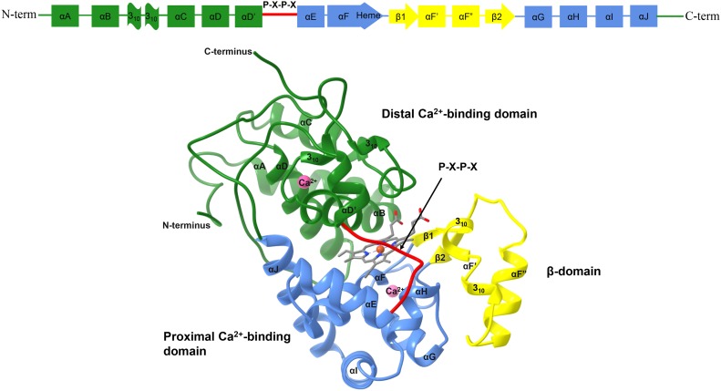
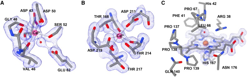
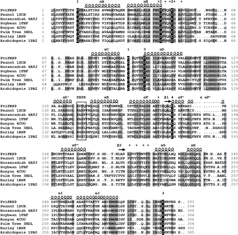
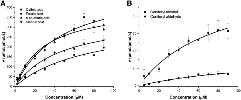
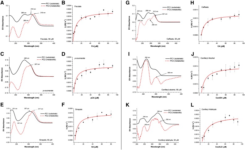
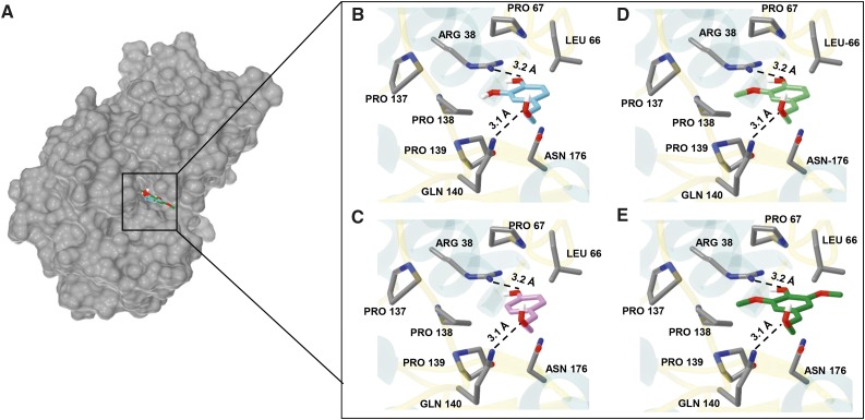
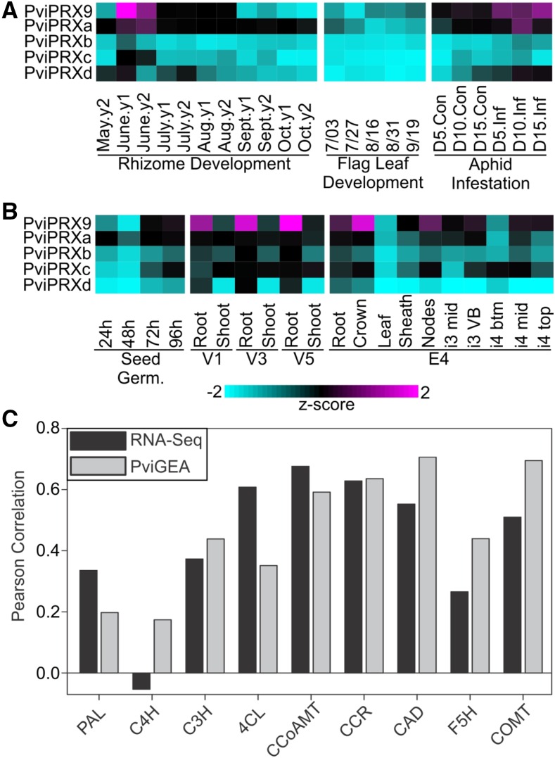
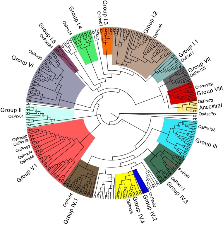
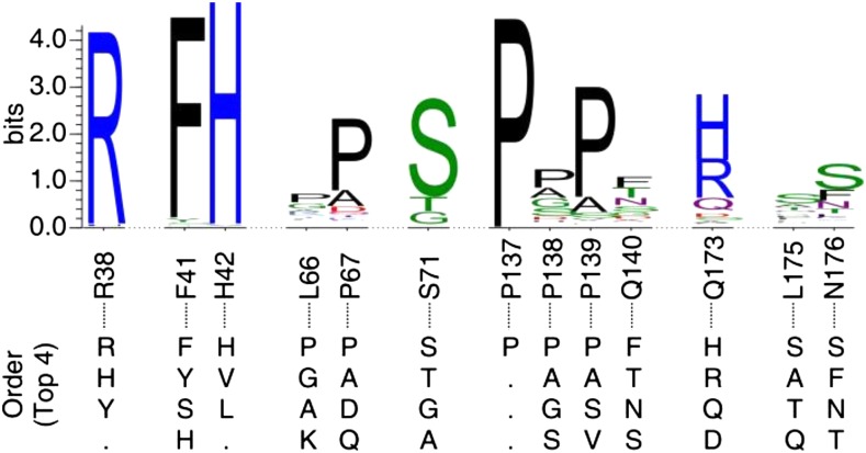
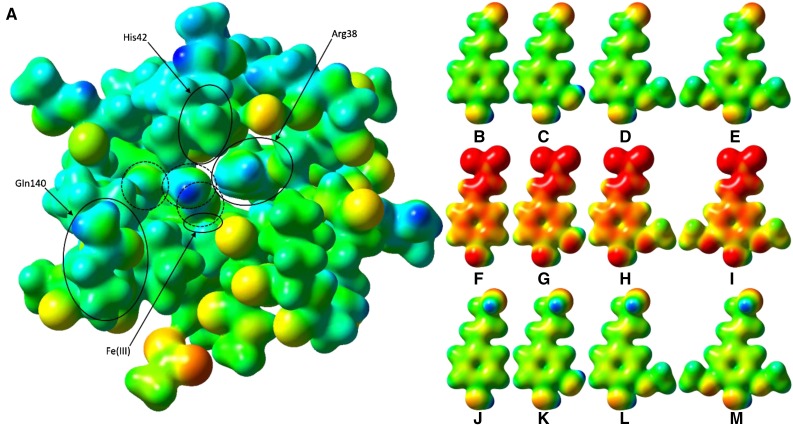
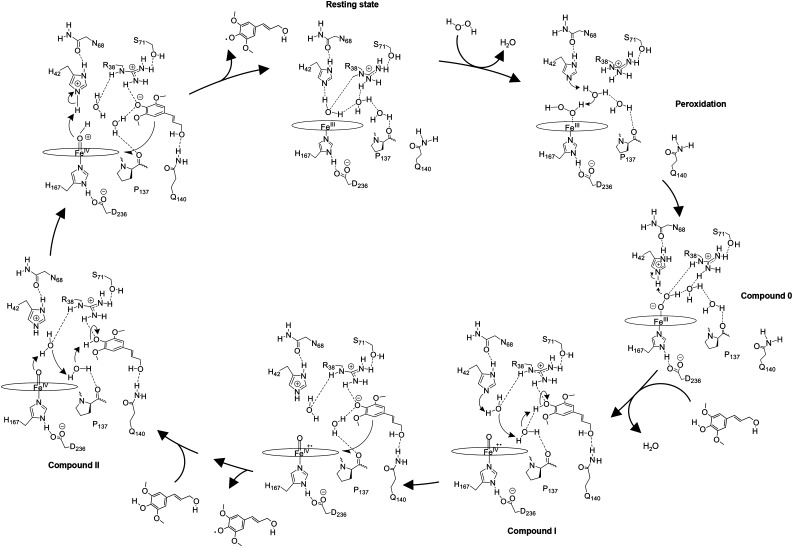
Class III peroxidases (CIIIPRX) catalyze the oxidation of monolignols, generate radicals, and ultimately lead to the formation of lignin. In general, CIIIPRX genes encode a large number of isozymes with ranges of in vitro substrate specificities. In order to elucidate the mode of substrate specificity of these enzymes, we characterized one of the CIIIPRXs (PviPRX9) from switchgrass (Panicum virgatum), a strategic plant for second-generation biofuels. The crystal structure, kinetic experiments, molecular docking, as well as expression patterns of PviPRX9 across multiple tissues and treatments, along with its levels of coexpression with the majority of genes in the monolignol biosynthesis pathway, revealed the function of PviPRX9 in lignification. Significantly, our study suggested that PviPRX9 has the ability to oxidize a broad range of phenylpropanoids with rather similar efficiencies, which reflects its role in the fortification of cell walls during normal growth and root development and in response to insect feeding. Based on the observed interactions of phenylpropanoids in the active site and analysis of kinetics, a catalytic mechanism involving two water molecules and residues histidine-42, arginine-38, and serine-71 was proposed. In addition, proline-138 and gluntamine-140 at the 137P-X-P-X140 motif, leucine-66, proline-67, and asparagine-176 may account for the broad substrate specificity of PviPRX9. Taken together, these observations shed new light on the function and catalysis of PviPRX9 and potentially benefit efforts to improve biomass conservation properties in bioenergy and forage crops.


Figures

Figure 1.
Figure 1.
Ribbon diagram representing the global structure of PviPRX9 with bound heme (gray) dividing the lower proximal Ca2+-binding domain (blue) containing the His-167 ligand, the upper distal Ca2+-binding domain (green), and the β-domain (yellow). The secondary structural elements were laid out as one-dimensional bars and are indicated following the convention of PRX. Ca2+ ions are depicted with pink balls. Molecular graphics images were produced using the Chimera package (Pettersen et al., 2004).
Figure 2.
Figure 2.
Ca2+ and heme iron coordination. A, The distal Ca2+ coordinated by the side chains of Asp-43, Asp-50, and Ser-52, backbone carbonyls of Val-46, Gly-48, and Asp-43, and a water molecule. B, The proximal Ca2+ coordinated by the side chains of Thr-168, Asp-211, Thr-214, and Asp-219 and the backbone carbonyls of Thr-168, Thr-214, and Thr-217. C, The active site showing the catalytic His-42, ligand-binding Arg-38, heme and the fifth heme iron ligand His-167, and the P-X-P-X motif of PviPRX9 (137PPPQ140). Images were rendered with Chimera version 1.11.
Figure 3.
Figure 3.
Structural alignment of PviPRX9 with seven CIIIPRXs from the Research Collaboratory for Structural Bioinformatics PDB. Secondary structural elements were labeled with helices indicated by spirals and β-sheets indicated by arrows. The secondary structure was letter mapped according to the system laid out by Schuller et al. (1996). Residues coordinating with the two conserved Ca2+ atoms are indicated with plus signs. The conserved P-X-P-X motif is outlined with a box. The Cys residues participating in conserved disulfides are numbered according to the corresponding Cys. Amino acids with 75% or greater identity in the alignment are highlighted in gray, and the conserved Cys residues are highlighted in black.
Figure 4.
Figure 4.
Steady-state initial rates are plotted versus reducing substrate concentrations. A, Carboxylates were varied from 2 to 90 µm, and the concentration of H2O2 was held constant at 500 µm. B, Coniferyl alcohol and coniferyl aldehyde were varied from 5 to 90 µm, and the concentration of H2O2 was held constant at 500 µm. Plots were generated using OriginPro 2016, and final graphs were generated with Microsoft Excel 2015.
Figure 5.
Figure 5.
Metabolite kinetics of PviPRX9. A, Plots of the ferulate substrate spectrum and the product spectrum showing a clear overlap of wavelengths. B, Michaelis-Menten plot for ferulate product formation. C, Plots of the p-coumarate substrate spectrum and the product spectrum showing wavelength overlap. D, Michaelis-Menten plot for p-coumarate product formation. E, Plots of the sinapate substrate spectrum and the product spectrum showing wavelength overlap. F, Michaelis-Menten plot for sinapate product formation. G, Plots of the caffeate substrate spectrum and the product spectrum showing wavelength overlap. H, Michaelis-Menten plot for caffeate product formation. I, Plots of the coniferyl alcohol substrate spectrum and the product spectrum showing wavelength overlap. J, Michaelis-Menten plot for coniferyl alcohol product formation. K, Plots of the coniferyl aldehyde substrate spectrum and the product spectrum showing wavelength overlap. L, Michaelis-Menten plot for coniferyl aldehyde product formation. The Michaelis-Menten metabolite plot y axis units are arbitrary units (AU) per second. Plots were generated using SVD/ALS with Mathcad and OriginPro 2016.
Figure 6.
Figure 6.
Active site of PviPRX9 with docked substrates coniferyl alcohol, p-coumaryl alcohol, sinapyl alcohol, and caffeyl alcohol. A, Surface representation of PviPRX9 showing the binding pocket with docked monolignols. B, Caffeyl alcohol is pictured in cyan oriented with the meta phenolic oxygen 3.2 Å from Arg-38 Nη2, and the propenyl oxygen was 3.1 Å from the Nε2 of Gln-140. C, p-Coumaryl alcohol is pictured in purple docked with phenolic oxygen oriented 3.2 Å from Arg-38 Nη2, and the propenyl oxygen was 3.1 Å from the Nε2 of Gln-140. D, Coniferyl alcohol is shown in lime green docked with the phenolic oxygen position within the hydrogen-bonding distance of 3.2 Å from Arg-38 Nη2, and the propenyl oxygen was 3.1 Å from the Nε2 of Gln-140. E, Sinapyl alcohol (forest green) docked with the phenolic oxygen 3.2 Å from Arg-38 Nη2, and the propenyl oxygen was 3.1 Å from the Nε2 of Gln-140. Molecular graphics images were produced using the Chimera package (Pettersen et al., 2004).
Figure 7.
Figure 7.
PviPRX9 expression analysis and relationship to the expression levels of genes associated with lignin biosynthesis. A, RNA-Seq-based transcript abundances of PviPRX9 and four additional peroxidases with correlated expression (PviPRXa to PviPRXd) in diverse switchgrass tissues shown as z-scores. Data are shown for rhizome development based on RNA-Seq experiments with rhizomes collected from field-grown plants of cv Summer over 2 years; flag leaf development; and aphid infestation with RNA-Seq analysis of tissues collected from uninfested (Con) and aphid-infested (Inf) cv Summer plants at 5, 10, and 15 d (D) post infestation. B, Microarray-based transcript abundances from PviGEA for PviPRX9 and four additional peroxidases with correlated expression (PviPRXa to PviPRXd) in multiple tissues and developmental stages shown as z-scores. Data are shown for four time points during seed germination (Seed Germ.); root and shoot tissue at three vegetative growth stages (V1, V3, and V5); and multiple tissues at the stem elongation 4 (E4) stage: root, crown, leaf (pooled leaves from E4 tiller), sheath (pooled sheaths from E4 tiller), nodes (pooled nodes from E4 tiller), the middle of the third internode (i3 mid), vascular bundles from the third internode (i3 VB), bottom one-fifth of the fourth internode (i4 btm), middle one-fifth of the fourth internode (i4 mid), and top one-fifth of the fourth internode (i4 top). Respective data sets and bioinformatic routines used for these analyses are given in “Materials and Methods.” Magenta indicates high expression and cyan indicates low expression. C, Correlation of transcript abundances between PviPRX9 and genes associated with monolignol biosynthesis in the data sets described in A and B. PAL, Phe ammonia lyase; C4H, cinnamate 4-hydroxylase; C3H, p-coumarate 3-hydroxylase; 4CL, 4-coumarate:CoA ligase; CCoAMT, caffeoyl-CoA o-methyltransferase; CCR, cinnamoyl-CoA reductase; CAD, cinnamoyl alcohol dehydrogenase; F5H, ferulate 5-hydroxylase; COMT, caffeic acid o-methyltransferase.
Figure 8.
Figure 8.
Maximum likelihood tree of all full-length CIIIPRXs in switchgrass. Maximum likelihood analysis was performed using the program Garli with 500 bootstrap pseudoreplicates. PviPRXs were assigned to evolutionary groups based on a phylogenetic grouping with previously assigned rice peroxidases. Dots indicate nodes with bootstrap support of 50 or greater. PviPRX9 was allocated in group IV.3 with the node in red.
Figure 9.
Figure 9.
Sequence logos of the residues constituting the substrate-binding pocket in all switchgrass CIIIPRXs. The top four amino acids at each position for all switchgrass CIIIPRXs were included below at their matching positions. Position labels were based on the sequence of PviPRX9. This figure was generated by WebLogo 3 (http://weblogo.threeplusone.com). The distributions of amino acids found in all positions are given in Supplemental Table S2.
Figure 10.
Figure 10.
Electrostatic potential surfaces of PviPRX9 and phenylpropanoids. A, Heme (hexcoordinate, high spin), Arg-31 (as methylguanidinium), Ser-35 (as ethanol), Arg-38 (as n-propylguanidinium), Phe-41 (as toluene), His-42 (as 5-methyl-1H-imidazole; mostly obscured by Leu-66), Ala-65 (as ACE) to Arg-73 (as NMA), Gly-170 (as formamide), Asp-135 (as ACE) to Phe-141 (as NMA), Ala-172 (as ACE) to Phe-177 (as NMA), His-167 (as 5-methyl-1H-imidazole), Asp-326 (as acetate ion), and six water molecules; Cys-174 was mutated to Gly to reduce computational cost. ACE and NMA are acetyl and N-methyl capping groups on the N and C termini, respectively, of each stretch of multiple residues. The three water molecules circled with dashed lines were, from right to left, the water involved in the first proton transfer, a noncatalytic water that occupies the nascent binding site of the hydroxycinnamyl substrate phenol functional group, and the water released upon H2O2 binding to the heme. The latter water molecule was regenerated by heterolytic cleavage of H2O2 and involved in catalytic proton transfer. B, p-Coumaryl aldehyde. C, Caffeyl aldehyde. D, Coniferyl aldehyde. E, Sinapyl aldehyde. F, p-Coumarate. G, Caffeate. H, Ferulate. I, Sinapate. J, p-Coumaryl alcohol. K, Caffeyl alcohol. L, Coniferoyl alcohol. M, Sinapyl alcohol. The active site and ligand electrostatic potentials, mapped on their corresponding self-consistent field densities, are shown on a potential scale of −2.7 × 10⁻¹ hartrees (red) to +2.97 × 101 hartrees (blue) for the active site and −1 × 10⁻¹ (red) to 3 × 101 (blue) for the ligands. This figure was generated using GaussView 3.09.
Figure 11.
Figure 11.
Proposed catalytic reaction mechanism of PviPRX9. In the first step, H2O2 displaces a water molecule from the active site in its resting state and reacts with His-42 to generate a hydroperoxide molecule via a water-mediated proton transfer. Next, the hydroperoxide molecule coordinates Fe(III) heme to generate Fe(III)-hydroperoxo heme (compound 0). In the second step, the hydroperoxo ligand is protonated by His-42 to generate first water and an Fe(IV)-oxo heme π-cation radical (compound I). Then, a monolignol binds to PviPRX9, displacing a water. His-42 deprotonates the monolignyl 4-hydroxy through a water-mediated proton-shuttling mechanism, generating the negatively charged monolignol that donates an electron to compound I to generate the first monolignol radical and Fe(IV)-oxo heme (compound II). In the third step, a second monolignol binds and is deprotonated through a water-mediated proton shuttle. Next, an electron is transferred to the heme from the deprotonated monolignol and His-42 protonates the Fe(IV)-hydroxo heme, generating the second water molecule along with the second monolignol radical, which diffuses from the pocket, returning PviPRX9 to its resting state.

References

    1. Abelskov AK, Smith AT, Rasmussen CB, Dunford HB, Welinder KG (1997) pH dependence and structural interpretation of the reactions of Coprinus cinereus peroxidase with hydrogen peroxide, ferulic acid, and 2,2′-azinobis(3-ethylbenzthiazoline-6-sulfonic acid). Biochemistry 36: 9453–9463 - PubMed
    1. Adams PD, Afonine PV, Bunkóczi G, Chen VB, Davis IW, Echols N, Headd JJ, Hung LW, Kapral GJ, Grosse-Kunstleve RW, et al. (2010) PHENIX: a comprehensive Python-based system for macromolecular structure solution. Acta Crystallogr D Biol Crystallogr 66: 213–221 - PMC - PubMed
    1. Almagro L, Gómez Ros LV, Belchi-Navarro S, Bru R, Ros Barceló A, Pedreño MA (2009) Class III peroxidases in plant defence reactions. J Exp Bot 60: 377–390 - PubMed
    1. Baek HK, Van Wart HE (1989) Elementary steps in the formation of horseradish peroxidase compound I: direct observation of compound 0, a new intermediate with a hyperporphyrin spectrum. Biochemistry 28: 5714–5719 - PubMed
    1. Bakovic M, Dunford HB (1993) Kinetics of the oxidation of p-coumaric acid by prostaglandin H synthase and hydrogen peroxide. Biochemistry 32: 833–840 - PubMed

Publication types

MeSH terms

LinkOut - more resources