Review
. 2016 Dec 5;215(5):611-619.
doi: 10.1083/jcb.201607062. Epub 2016 Nov 22.

Meiotic spindle assembly and chromosome segregation in oocytes

Affiliations
Review

Meiotic spindle assembly and chromosome segregation in oocytes

Isma Bennabi et al. J Cell Biol. .

Abstract

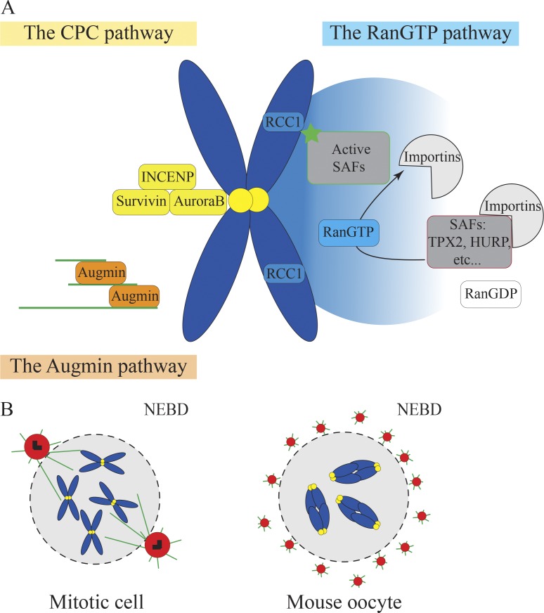
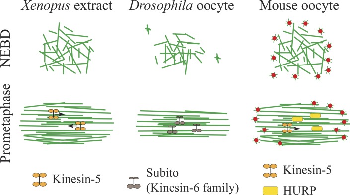
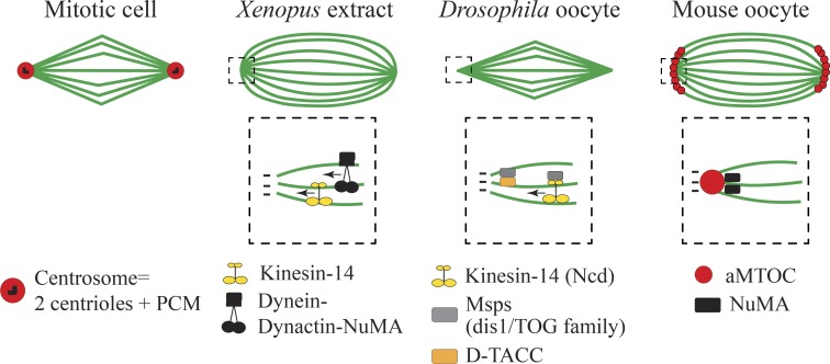
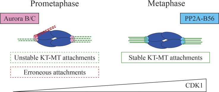
Oocytes accumulate maternal stores (proteins, mRNAs, metabolites, etc.) during their growth in the ovary to support development after fertilization. To preserve this cytoplasmic maternal inheritance, they accomplish the difficult task of partitioning their cytoplasm unequally while dividing their chromosomes equally. Added to this complexity, most oocytes, for reasons still speculative, lack the major microtubule organizing centers that most cells use to assemble and position their spindles, namely canonical centrosomes. In this review, we will address recent work on the mechanisms of meiotic spindle assembly and chromosome alignment/segregation in female gametes to try to understand the origin of errors of oocyte meiotic divisions. The challenge of oocyte divisions appears indeed not trivial because in both mice and humans oocyte meiotic divisions are prone to chromosome segregation errors, a leading cause of frequent miscarriages and congenital defects.


Figures

Figure 1.
Figure 1.
Pathways replacing centrosomes for microtubule nucleation in oocytes. (A) The three microtubule nucleation pathways: the RanGTP pathway, CPC pathway, and Augmin pathway. (B) Microtubule nucleation by centrosomes in mitotic cells (left) and by multiple aMTOCs in mouse oocytes (right). For A and B, DNA is in blue, microtubules in green, kinetochores in yellow, pericentriolar material in red, and centrioles in black.
Figure 2.
Figure 2.
Spindle bipolarization. Organization of microtubules into a bipolar array via microtubule motors and microtubule-associated proteins in Xenopus egg extracts, Drosophila, and mouse oocytes between NEBD and prometaphase. Microtubules are in green and aMTOCs in red.
Figure 3.
Figure 3.
Spindle pole formation and final spindle shape. Top rows show spindle shape in metaphase in mitotic cells, Xenopus egg extracts, Drosophila, and mouse oocytes. The dashed square shows magnification of the spindle pole where microtubule motors and microtubule-associated proteins organize microtubule minus ends. Microtubules are in green.
Figure 4.
Figure 4.
Establishment of stable kinetochoremicrotubule attachments in mouse oocytes. Mouse oocytes form stable kinetochore–microtubule (KT-MT) attachments only at late metaphase I. Aurora B/C phosphorylation destabilizes kinetochore–microtubule attachments, whereas PP2A-B56 dephosphorylation activity stabilizes the attachments. This process is regulated by a progressive increase in CDK1 activity. DNA is in blue.

References

    1. Alvarez Sedó C., Schatten H., Combelles C.M., and Rawe V.Y.. 2011. The nuclear mitotic apparatus (NuMA) protein: Localization and dynamics in human oocytes, fertilization and early embryos. Mol. Hum. Reprod. 17:392–398. 10.1093/molehr/gar009 - DOI - PubMed
    1. Azimzadeh J., Wong M.L., Downhour D.M., Sánchez Alvarado A., and Marshall W.F.. 2012. Centrosome loss in the evolution of planarians. Science. 335:461–463. 10.1126/science.1214457 - DOI - PMC - PubMed
    1. Balboula A.Z., and Schindler K.. 2014. Selective disruption of aurora C kinase reveals distinct functions from aurora B kinase during meiosis in mouse oocytes. PLoS Genet. 10:e1004194 10.1371/journal.pgen.1004194 - DOI - PMC - PubMed
    1. Basto R., Lau J., Vinogradova T., Gardiol A., Woods C.G., Khodjakov A., and Raff J.W.. 2006. Flies without centrioles. Cell. 125:1375–1386. 10.1016/j.cell.2006.05.025 - DOI - PubMed
    1. Bayliss R., Sardon T., Vernos I., and Conti E.. 2003. Structural basis of Aurora-A activation by TPX2 at the mitotic spindle. Mol. Cell. 12:851–862. 10.1016/S1097-2765(03)00392-7 - DOI - PubMed

LinkOut - more resources