Review
. 2016 Nov 22;17(11):1949.
doi: 10.3390/ijms17111949.

Structure-Functional Basis of Ion Transport in Sodium-Calcium Exchanger (NCX) Proteins

Affiliations
Review

Structure-Functional Basis of Ion Transport in Sodium-Calcium Exchanger (NCX) Proteins

Moshe Giladi et al. Int J Mol Sci. .

Abstract

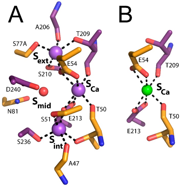
The membrane-bound sodium-calcium exchanger (NCX) proteins shape Ca2+ homeostasis in many cell types, thus participating in a wide range of physiological and pathological processes. Determination of the crystal structure of an archaeal NCX (NCX_Mj) paved the way for a thorough and systematic investigation of ion transport mechanisms in NCX proteins. Here, we review the data gathered from the X-ray crystallography, molecular dynamics simulations, hydrogen-deuterium exchange mass-spectrometry (HDX-MS), and ion-flux analyses of mutants. Strikingly, the apo NCX_Mj protein exhibits characteristic patterns in the local backbone dynamics at particular helix segments, thereby possessing characteristic HDX profiles, suggesting structure-dynamic preorganization (geometric arrangements of catalytic residues before the transition state) of conserved α₁ and α₂ repeats at ion-coordinating residues involved in transport activities. Moreover, dynamic preorganization of local structural entities in the apo protein predefines the status of ion-occlusion and transition states, even though Na⁺ or Ca2+ binding modifies the preceding backbone dynamics nearby functionally important residues. Future challenges include resolving the structural-dynamic determinants governing the ion selectivity, functional asymmetry and ion-induced alternating access. Taking into account the structural similarities of NCX_Mj with the other proteins belonging to the Ca2+/cation exchanger superfamily, the recent findings can significantly improve our understanding of ion transport mechanisms in NCX and similar proteins.

Keywords: HDX-MS (hydrogen–deuterium exchange mass-spectrometry); NCX (sodium–calcium exchanger); alternating access; antiporter; catalysis; occlusion; selectivity; transporter.


Conflict of interest statement

The authors declare no conflict of interest.

Figures

Figure 1
Figure 1
Structure of NCX_Mj (Methanococcus jannaschii sodium–calcium exchanger). (A) crystal structure of outward-facing NCX_Mj (PDB 3V5U) in cartoon representation. Helices 1–5 (TM1–5) are orange and helices 6–10 (TM6–10) are purple. Purple and green spheres represent Na+ and Ca2+ ions, respectively; (B) crystal structure of inward-facing VCX1 (PDB 4K1C) in cartoon representation. Helices 1–5 (TM1–5) are light orange and helices 6–10 (TM6–10) are magenta. Green sphere represents Ca2+; and (C) ion coordination as initially suggested by the crystal structure of NCX_Mj (PDB 3V5U). Ion coordinating residues are shown as sticks. Purple and green spheres represent Na+ and Ca2+ ions, respectively.
Figure 2
Figure 2
Ion binding sites of NCX_Mj. (A) Na+ binding sites (PDB 5HXE); (B) Ca2+ binding site (PDB 3V5U). Ion coordinating residues are presented as sticks. Purple and green spheres represent Na+ and Ca2+ ions, respectively.
Figure 3
Figure 3
Ion transport cycle and NCX backbone dynamics. (A) schematic representation of the ion-flux assay for the Na+/45Ca2+ exchange or the Ca2+/45Ca2+ exchange and the ping-pong mechanism describing the exchange reactions. The red, green, and purple arrows represent Ca2+-entry, Ca2+-exit, and Na+-exit steps of the transport cycle, respectively. The dotted arrows represent the reactions of the transport cycle, which negligibly contribute to the observed ion-exchange reactions under the given experimental conditions. The K’d and K”d values represent the dissociation constants for Ca2+ binding to NCX_Mj at the cytosolic and extracellular sides, respectively; and (BD) the heat map after a 1200 s exchange is overlaid on the crystal structure of NCX_Mj for the apo (PDB 5HXH, B), Na+-bound (PDB 5HXE, C), and Ca2+-bound (PDB 5HXR, D) forms. The color key indicates the HDX level. The numbers indicate the transmembrane helix number.
Figure 4
Figure 4
Ligand-induced alternating access in NCX_Mj. (A) the crystal structures of NCX_Mj bound to 3Na+ ions (blue, PDB 5HXE) and 2Na+ ions (gray, PDB 5HWX) are superimposed and viewed from the membrane plane (left) and from the extracellular side (right). Note the change in distance between TM6 and TM7ab induced by ligand binding; (B) the proposed “one-transition/two-occluded state” model for Ca2+ translocation from the extracellular to the cytosolic side, whereas the Ca2+-occluded state is more stable in the extracellular orientation (the outward-facing state) as compared with the cytosolic one (the inward-facing state). [E·Ca] is the Ca2+-bound transition state; and (C) the Na+/Ca2+ exchange cycle is described as a separate translocation of 1Ca2+ or 3Na+, where the extracellular occlusion of either ion is more stable than the cytosolic one. Green and red spheres represent Na+ and Ca2+ ions, respectively. The gating bundle (TM1/TM6) is represented as a dashed line. In the frame of the “one-transition/two-occluded state” model, the movement of the TM1/TM6 bundle is driven by the transition state and not by the occlusion or semi-open states.
Figure 5
Figure 5
Ion selectivity of NCX. (A) sequence alignment of the α-repeats of NCX_Mj and members of the Na+/Ca2+-K+ exchanger (NCKX), Ca2+/H+ exchanger (CAX) and mitochondrial Na+/Ca2+ exchanger (NCLX) families. Ion-coordinating residues are red; (B) 3Na+ ions (purple spheres, 5HXE) coordination and 1Ca2+ ion (green sphere, 5HXR) coordination as suggested by the crystal structures of NCX_Mj. Mutated residues in the NCX_Mj/NCLX construct are red.

References

    1. Roux B., Berneche S., Egwolf B., Lev B., Noskov S.Y., Rowley C.N., Haibo Y. Ion selectivity in channels and transporters. J. Gen. Physiol. 2010;137:415–426. doi: 10.1085/jgp.201010577. - DOI - PMC - PubMed
    1. Dudev T., Lim C. Ion selectivity strategies of sodium channel selectivity filters. Chem. Rev. 2014;114:538–556. doi: 10.1021/cr4004665. - DOI - PubMed
    1. Lockless S.W. Determinants of cation transport selectivity: Equilibrium binding and transport kinetics. J. Gen. Physiol. 2015;146:3–13. doi: 10.1085/jgp.201511371. - DOI - PMC - PubMed
    1. Carafoli E. Intracellular calcium homeostasis. Annu. Rev. Biochem. 1987;56:395–433. doi: 10.1146/annurev.bi.56.070187.002143. - DOI - PubMed
    1. Berridge M.J., Bootman M.D., Roderick H.L. Calcium signalling: Dynamics, homeostasis and remodelling. Nat. Rev. Mol. Cell Biol. 2003;4:517–529. doi: 10.1038/nrm1155. - DOI - PubMed

MeSH terms

LinkOut - more resources