Review
. 2008 Apr 17;8(4):2722-2735.
doi: 10.3390/s8042722.

Visible Genotype Sensor Array

Affiliations
Review

Visible Genotype Sensor Array

Yuichi Michikawa et al. Sensors (Basel). .

Abstract

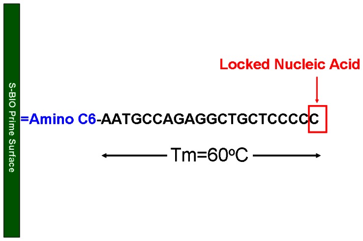
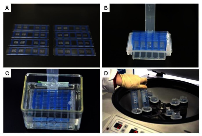
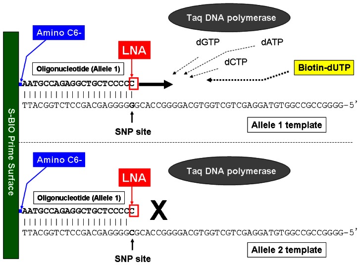
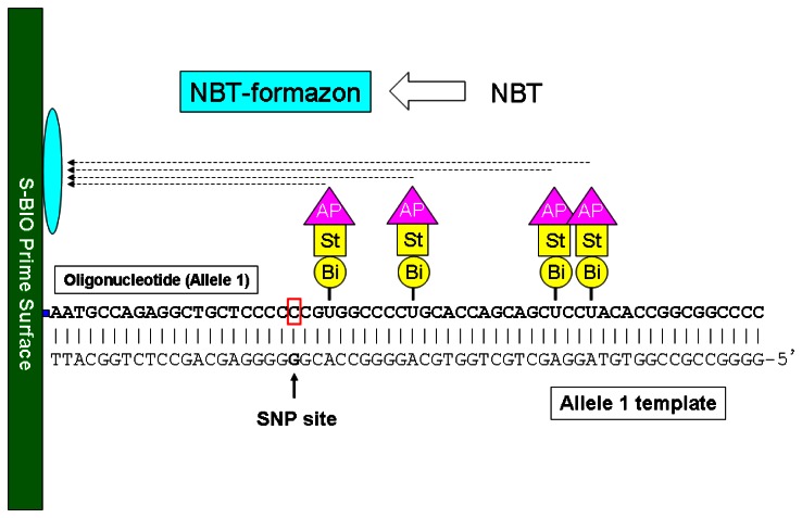
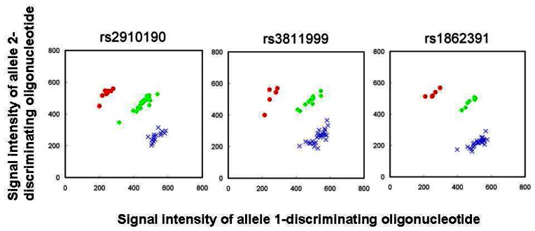
A visible sensor array system for simultaneous multiple SNP genotyping has been developed using a new plastic base with specific surface chemistry. Discrimination of SNP alleles is carried out by an allele-specific extension reaction using immobilized oligonucleotide primers. The 3'-ends of oligonucleotide primers are modified with a locked nucleic acid to enhance their efficiency in allelic discrimination. Biotin-dUTPs included in the reaction mixture are selectively incorporated into extending primer sequences and are utilized as tags for alkaline phosphatase-mediated precipitation of colored chemical substrates onto the surface of the plastic base. The visible precipitates allow immediate inspection of typing results by the naked eye and easy recording by a digital camera equipped on a commercial mobile phone. Up to four individuals can be analyzed on a single sensor array and multiple sensor arrays can be handled in a single operation. All of the reactions can be performed within one hour using conventional laboratory instruments. This visible genotype sensor array is suitable for "focused genomics" that follows "comprehensive genomics".

Keywords: SNP; Visible sensor; array; plastic; primer extension..


Figures

Figure 1.
Figure 1.
Recording the visible genotype sensor array image using a digital camera equipped on a mobile phone. A: Image recording using a mobile phone. B: Recorded image on the mobile phone. Individual spots on the sensor array indicate target SNP allele types. Visibility of the genotyping spots allows immediate inspection of results.
Figure 2.
Figure 2.
Structure of an allele-discriminating immobilized oligonucleotide. The 3′-end nucleotide opposing the target SNP nucleotide in the template DNA is LNA modified to enhance allelic discrimination efficiency. The Tm of the backbone oligonucleotide is set to be 60°C. Amino C6 is attached to the 5′-end nucleotide for covalent immobilization to the surface of a plastic base.
Figure 3.
Figure 3.
Spotting of oligonucleotides on plastic bases. A: MassARRAY Nanodispensor. B: Spotting on a plastic base. C: Heating of the spotted plastic base. D: Comparison of original and spotted plastic bases.
Figure 4.
Figure 4.
Post spotting processes in preparation of visible genotype sensor array. A: Attaching a Multiwell Geneframe onto each plastic base. B: Simultaneous handling of multiple plastic bases. C: Masking unspotted surface of plastic bases by soaking in 1 x S-BIO blocking solution. D: Removal of surface solution by centrifugation.
Figure 5.
Figure 5.
Scheme of reaction processes on the visible genotype sensor array. Only three reaction processes are necessary. These reactions can be performed by conventional laboratory instruments shown in Figure 9. Typing results can be visibly inspected within one hour.
Figure 6.
Figure 6.
Allele-specific primer extension reaction. The immobilized oligonucleotide (Allele 1) that is hybridized to perfectly matched template (Allele 1 template) can be extended according to the sequence of the template. Biotin-dUTPs in the solution are thus incorporated during the extension process. Extension of the immobilized oligonucleotide (Allele 1) hybridized to a mismatched template (Allele 2 template) is efficiently inhibited by the LNA modification at its 3′ end.
Figure 7.
Figure 7.
Binding of alkaline phosphatase-conjugated streptavidin to the incorporated biotin-dUTPs. Streptavidin specifically binds to biotin molecules in the extended sequence of the immobilized oligonucleotide. This reaction allows spatially restricted localization of the alkaline phosphatase-streptavidin conjugates on the plastic base.
Figure 8.
Figure 8.
Colored substrate precipitation. Captured alkaline phosphatases catalyze conversion of soluble NBT into unsoluble, colored NBT-formazon. The resulting NBT-formazon precipitate adheres to the surface of the plastic base, giving visible spots.
Figure 9.
Figure 9.
Overview of experimental set-up for reactions on the sensor array. A: Set-up of allele-specific primer extension. The reaction mixture was added to each well of the visible genotype sensor arrays then placed on a plastic tip case containing pre-warmed water. B: Allele-specific primer extension was performed in a constant temperature incubator at 65°C. The plastic tip case was wrapped with Saran wrap to preserve humidity. C: Incubation of alkaline phosphatase-conjugated streptavidin. D: Color development using BCIP/NBT.
Figure 10.
Figure 10.
Examples of spot images from the visible genotype sensor array. Genotypes of four individuals were simultaneously analyzed on one sensor array. Allele 1: Allele-1 disciminating oligonucleotides spotted in triplicate. Allele 2: Allele-2 discriminating oligonucleotides spotted in triplicate. P: Positive control oligonucleotide [16]. N: Negative control oligonucleotide [16].
Figure 11.
Figure 11.
Scatter plots of signal intensities measured for three SNPs. X-axis: Signal intensity of allele-1 discriminating oligonucleotide. Y-axis: Signal intensity of allele-2 discriminating oligonucleotide. Data plots were of 45 individuals previously genotyped by another established method (MassARRAY). Blue crosses: Allele 1 homozygotes determined by MassARRAY. Green open diamonds: Heterozygotes determined by MassARRAY. Red closed circles: Allele 2 homozygotes determined by MassARRAY. Plots are modified from ref. .

References

    1. Kruglyak L. Prospects for whole-genome linkage disequilibrium mapping of common disease genes. Nature Genet. 1999;22:139–144. - PubMed
    1. Ohashi J., Tokunaga K. The power of genome-wide association studies of complex disease genes: statistical limitations of indirect approaches using SNP markers. J. Hum. Genet. 2001;46:478–482. - PubMed
    1. Wang W.Y., Barratt B.J., Clayton D.G., Todd J.A. Genome-wide association studies: theoretical and practical concerns. Nature Rev. Genet. 2005;6:109–118. - PubMed
    1. Thomas D.C. Are we ready for genome-wide association studies? Cancer Epidemiol. Biomark. Prev. 2006;15:595–598. - PubMed
    1. Klein R.J. Power analysis for genome-wide association studies. BMC Genet. 2007;8:58. - PMC - PubMed