. 2008 Jul 28;8(7):4413-4428.
doi: 10.3390/s8074413.

Nanobioengineering and Characterization of a Novel Estrogen Receptor Biosensor

Affiliations

Nanobioengineering and Characterization of a Novel Estrogen Receptor Biosensor

Alexandre Berthier et al. Sensors (Basel). .

Abstract

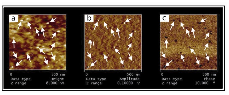
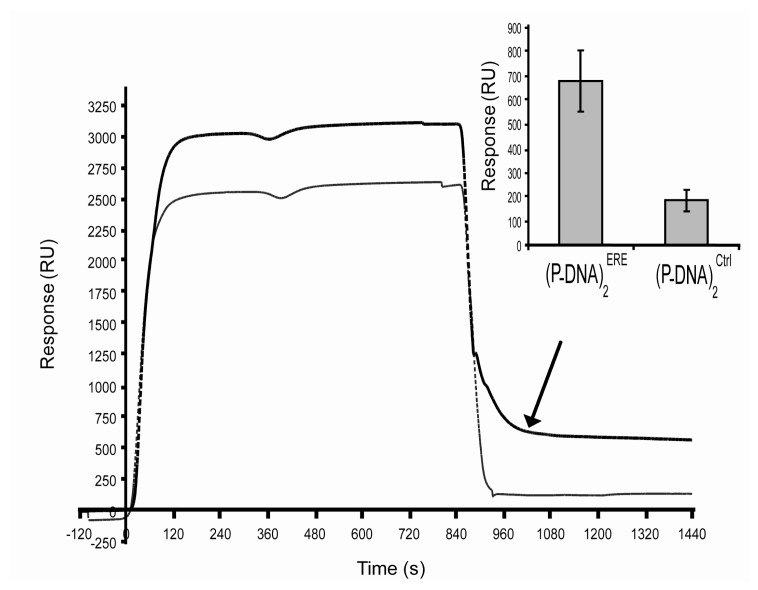
We constructed an original supramolecular assembly on a surface of sensor composed of an innovative combination of an engineered cytochrome b5 and a modified nucleic acid bound to a synthetic lipid hemimembrane. The protein/DNA block, called (PDNA) 2, was synthesized and purified before its immobilization onto a hybrid bilayer reconstituted on a gold surface. Surface plasmon resonance (SPR) and atomic force microscopy (AFM) were engaged in parallel on the same substrates in order to better understand dynamic events that occur at the surface of the biosensor. Good correlations were obtained in terms of specificity and reversibility. These findings allow us to present a first application of such biosensor in the study of the interaction processes between nuclear receptor and DNA.

Keywords: AFM; Nano-objects; Protein/DNA interaction.; SPR; molecular lego.


Figures

Figure 1.
Figure 1.
Specific assembling of (P-DNA)2onto the chip. (a) Sensorgram of the hybrid bilayer establishment. This HB was realized at 25°C, on functionalized gold surface in PB buffer. SAM was first wetted with 50% ethanol, and then washed with two pulses (1 min each) of 40 mM OG at 50 μl/min. 1 mM SUV (DOGS/DMPC 10% mol/mol) was injected at 2 μl/min and spread onto the cleaned surface. At the end of the injection, flow was increased to 20 μl/min and two pulses of 20 mM NaOH treated the lipidic surface in order to remove excess vesicles. DOGS were reloaded in Ni2+ by an injection of NiCl2 (20 mM in acetate buffer). At the end of this process, the response signal was ∼1700 RU corresponding to 240 pmol/cm2, (b) The sensorgram shows the control of the homogeneity of the HB through injections at 20 μl/min of a dummy protein (cytochrome C 1 μM). (c) Sensorgram of the (P-DNA)2Ctrl anchorage. This step was realized in PBS running buffer at 25°C at a concentration of 2 μM during 5 minutes at 5 μl/min. (d) Sensorgram showing the specific anchorage of the (P-DNA)2 block. The lipidic surface was regenerated by three pulses of 0.5 M imidazole (30 seconds at 20 μl/min).
Figure 2.
Figure 2.
Specificity and reversibility of the biosensor. AFM images of gold supported HB after DOGS reloading in Ni2+(a) after 20 min incubation of 200 nM cytochrome c (b), (P-DNA)2(c) and after imidazole incubation (d) obtained by contact mode imaging in liquid conditions. (e) Surface roughness (in nm) was determined on every image, on 1 and 4 mm2. z range corresponds to 15 nm in contact mode.
Figure 3.
Figure 3.
Visualization of (P-DNA)2 complexes immobilized onto the supported HB. The height (a) amplitude (b) and phase (c) representations from oscillating contact mode AFM images are nicely correlated after (P-DNA)2 incubation on the HB. White arrows indicate motifs. The supported membrane contains 1% nickel modified lipids and was reloaded with a 50 mM nickel solution in acetate buffer. The (P-DNA)2 complexes were incubated for 30 min onto the membrane, followed by extensive washes with PBS buffer.
Figure 4.
Figure 4.
Specific interaction between ERα and ERE target sequence. After 4 hours incubation of 50 nM ERα with 1 nM E2 in PBS at 4°C, 300 μl of “activated” receptors were injected at 20 μl/min on (P-DNA)2ERE (thick curve) or on (P-DNA)2Ctrl (thin curve). The graphic representation of the results was the mean of four experiments.

References

    1. Sackmann E. Supported membranes: scientific and practical applications. Science. 1996;271:43–48. - PubMed
    1. Tanaka M., Sackmann E. Polymer-supported membranes as models of the cell surface. Nature. 2005;437:656–663. - PubMed
    1. Larsson C., Bramfeldt H., Wingren C., Borrebaeck C., Hook F. Gravimetric antigen detection utilizing antibody-modified lipid bilayers. Anal. Biochem. 2005;345:72–80. - PubMed
    1. Suraniti E., Tumolo T., Baptista M.S., Livache T., Calemczuk R. Construction of hybrid bilayer membrane (HBM) Biochips and characterization of the cooperative binding between cytochrome-c and HBM. Langmuir. 2007;23:6835–6842. - PubMed
    1. Svedhem S., Dahlborg D., Ekeroth J., Kelly J., Höök F., Gold J. In Situ Peptide-Modified Supported Lipid Bilayers for Controlled Cell Attachment. Langmuir. 2003;19:6730–6736.

LinkOut - more resources