

Correlation of ultra-widefield fundus autofluorescence patterns with the underlying genotype in retinal dystrophies and retinitis pigmentosa
- PMID: 27880076
- PMCID: PMC6186264
- DOI: 10.1080/13816810.2016.1227450
Correlation of ultra-widefield fundus autofluorescence patterns with the underlying genotype in retinal dystrophies and retinitis pigmentosa
Abstract
Purpose: Ultra-widefield fundus autofluorescence (UW-FAF) allows the characterization of the peripheral retinal features of vitreoretinal diseases. The purpose of this study was to examine possible genotypic/phenotypic correlations of UW-FAF patterns in patients with a variety of retinal dystrophies and retinitis pigmentosa (RP).
Methods: An IRB-approved retrospective consecutive case series study was performed of genetically characterized retinal dystrophy or RP patients who underwent UW-FAF imaging. UW-FAF was performed with the Optos 200Tx system. Clinical variables, genotypic analysis, and phenotypic characteristics were reviewed.
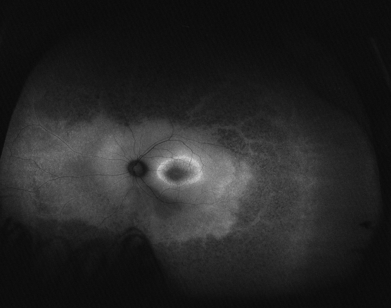
Results: Seventeen patients were identified who had identified mutations in retinal dystrophy or RP genes and who also had undergone UW-FAF. Three patients had X-linked RP with RPGR mutations. Six patients had autosomal dominant RP (four with RHO mutations and one with a PRPF31 mutation, and one with RDS/PRPH2 mutation). Four patients had autosomal recessive RP (four with USH2A mutations). Three patients had Leber Congenital Amaurosis (LCA) with mutations including CRB1, CEP290, and RPGRIP1. Macular hyperautofluorescence was noted in all patients. A ring of hyperautofluorescence was clear in patients with RHO and USH2A mutations, and patients with USH2A mutations demonstrated a second ring of hyperautofluorescence. In the periphery, patients with RHO or RPGR mutations exhibited hyperautofluorescence with patchy areas of hypoautofluorescence. Patients with USH2A mutations had a distinctive pattern of diffuse and homogeneous peripheral hypoautofluorescence.
Conclusion: UW-FAF may provide important information to facilitate diagnosis and further research is needed to better characterize this technology as an imaging biomarker for genotype association in retinal dystrophies and RP.
Keywords: Autofluorescence; fundus; retina.
Figures


References
-
- Haim M Epidemiology of retinitis pigmentosa in Denmark. Acta Ophthalmol. Scand Suppl. 80, 1–34 (2002). - PubMed
-
- Hartong DT1, Berson EL, Dryja TP. Retinitis pigmentosa. Lancet. 2006. November 18;368(9549):1795–809. - PubMed
-
- Heckenlively JR. Retinitis Pigmentosa. Philadelphia: Lippincott; 1988.
-
- Kaplan J, Bonneau D, Frezal J, Munnich A, Dufier J-L Clinical and genetic heterogeneity in retinitis pigmentosa. Hum. Genet 85: 635–642, 1990. - PubMed
-
- Gu S, Thompson DA, Srikumari CRS, Lorenz B, Finckh U, Nicoletti A, Murthy KR, Rathmann M, Kumaramanickavel G, Denton MJ, Gal A Mutations in RPE65 cause autosomal recessive childhood-onset severe retinal dystrophy. Nature Genet. 17: 194–197, 1997. - PubMed
Publication types
MeSH terms
Substances
Grants and funding
LinkOut - more resources
Full Text Sources
Other Literature Sources
Research Materials