. 2016 Nov 23;11(11):e0166984.
doi: 10.1371/journal.pone.0166984. eCollection 2016.

Wdr68 Mediates Dorsal and Ventral Patterning Events for Craniofacial Development

Affiliations

Wdr68 Mediates Dorsal and Ventral Patterning Events for Craniofacial Development

Estibaliz Alvarado et al. PLoS One. .

Abstract

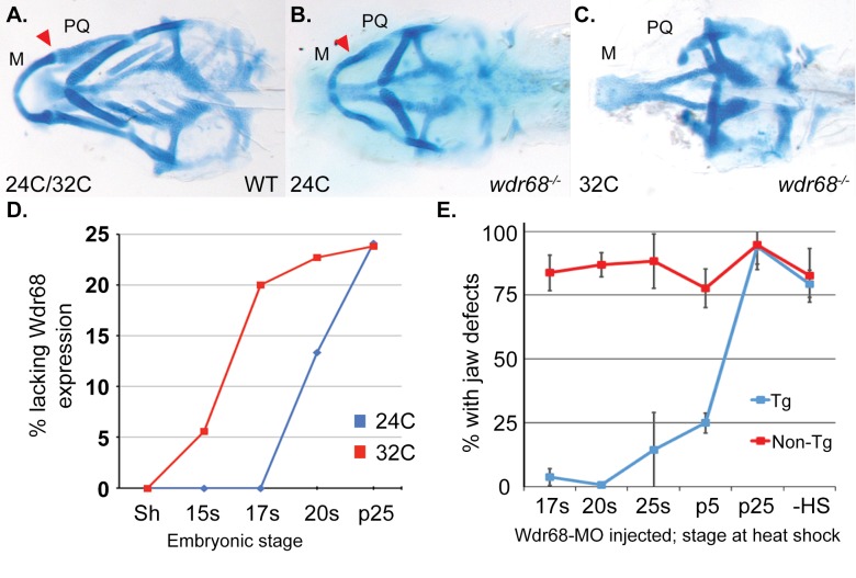
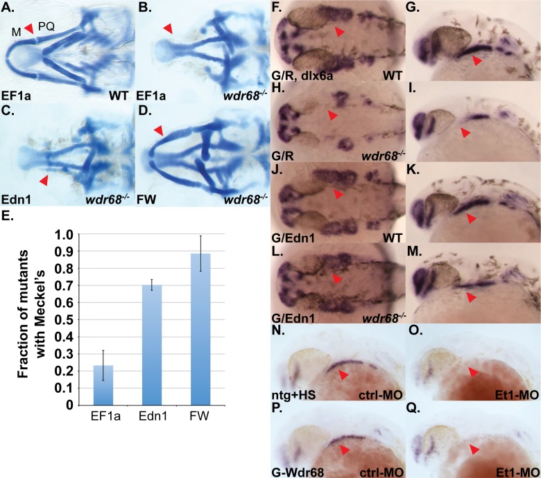
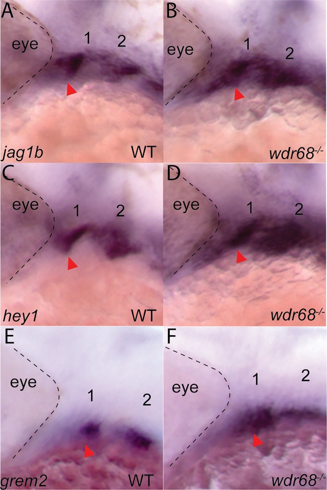
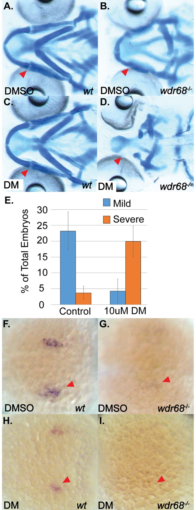
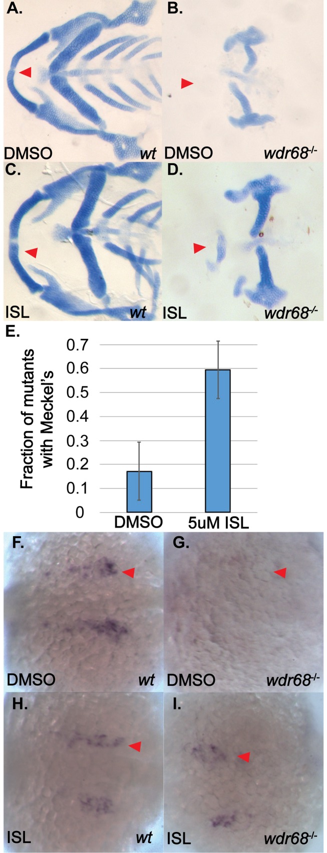
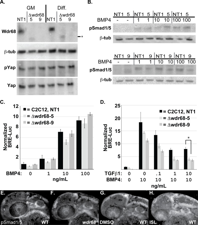
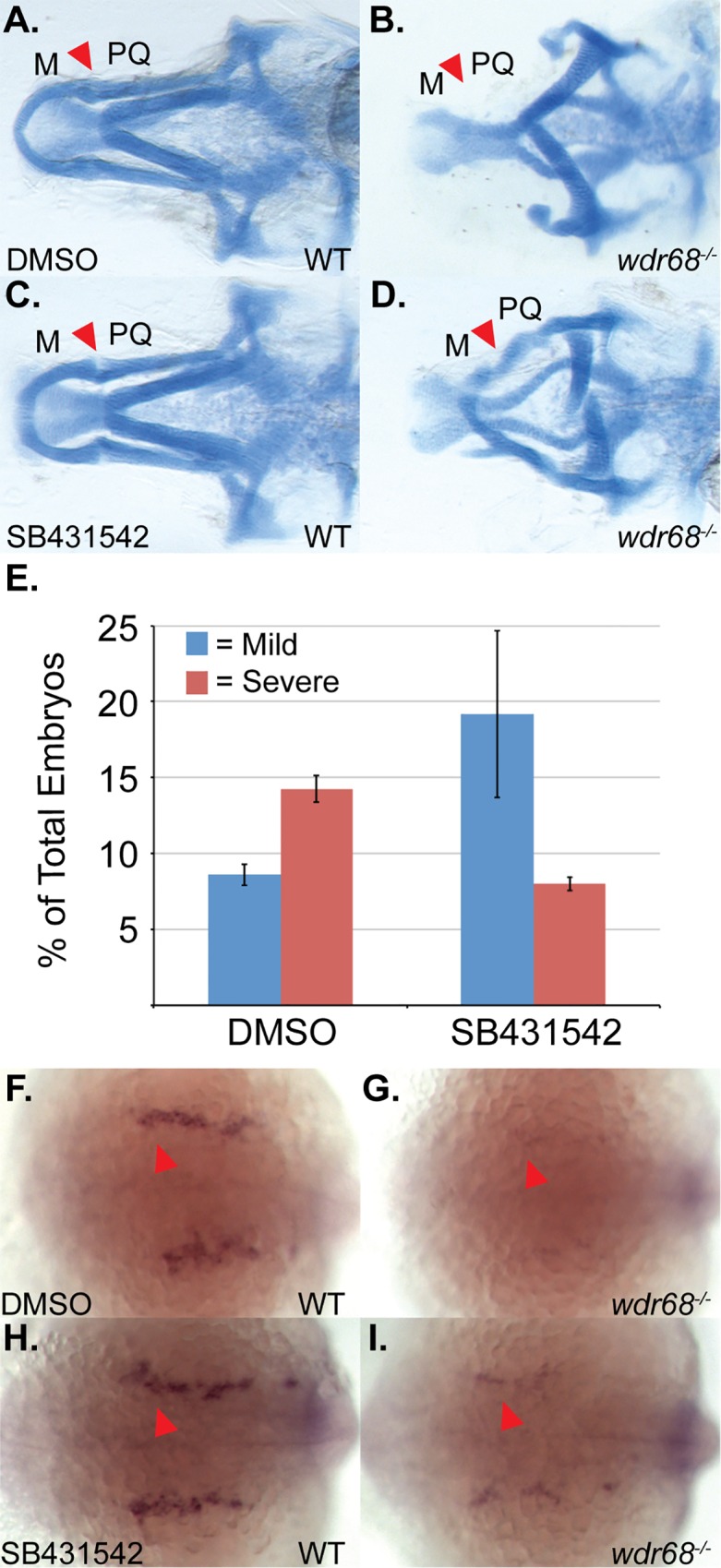
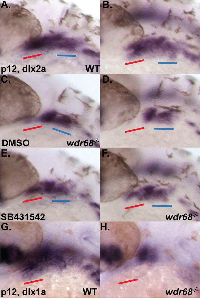
Birth defects are among the leading causes of infant mortality and contribute substantially to illness and long-term disability. Defects in Bone Morphogenetic Protein (BMP) signaling are associated with cleft lip/palate. Many craniofacial syndromes are caused by defects in signaling pathways that pattern the cranial neural crest cells (CNCCs) along the dorsal-ventral axis. For example, auriculocondylar syndrome is caused by impaired Endothelin-1 (Edn1) signaling, and Alagille syndrome is caused by defects in Jagged-Notch signaling. The BMP, Edn1, and Jag1b pathways intersect because BMP signaling is required for ventral edn1 expression that, in turn, restricts jag1b to dorsal CNCC territory. In zebrafish, the scaffolding protein Wdr68 is required for edn1 expression and subsequent formation of the ventral Meckel's cartilage as well as the dorsal Palatoquadrate. Here we report that wdr68 activity is required between the 17-somites and prim-5 stages, that edn1 functions downstream of wdr68, and that wdr68 activity restricts jag1b, hey1, and grem2 expression from ventral CNCC territory. Expression of dlx1a and dlx2a was also severely reduced in anterior dorsal and ventral 1st arch CNCC territory in wdr68 mutants. We also found that the BMP agonist isoliquiritigenin (ISL) can partially rescue lower jaw formation and edn1 expression in wdr68 mutants. However, we found no significant defects in BMP reporter induction or pSmad1/5 accumulation in wdr68 mutant cells or zebrafish. The Transforming Growth Factor Beta (TGF-β) signaling pathway is also known to be important for craniofacial development and can interfere with BMP signaling. Here we further report that TGF-β interference with BMP signaling was greater in wdr68 mutant cells relative to control cells. To determine whether interference might also act in vivo, we treated wdr68 mutant zebrafish embryos with the TGF-β signaling inhibitor SB431542 and found partial rescue of edn1 expression and craniofacial development. While ISL treatment failed, SB431542 partially rescued dlx2a expression in wdr68 mutants. Together these findings reveal an indirect role for Wdr68 in the BMP-Edn1-Jag1b signaling hierarchy and dorso-anterior expression of dlx1a/2a.


Conflict of interest statement

The authors have declared that no competing interests exist.

Figures

Fig 1
Fig 1. Wdr68 is required for craniofacial development between the 17 somites and prim-5 stages.
A) Ventral view of 5dpf alcian blue stained cartilages in wild type zebrafish. M: Meckel’s PQ: Palatoquadrate. B) Mild mutant phenotype resulting from rearing of embryos at 24°C, characterized by joint fusions (arrowhead) between M and PQ. C) Severe mutant phenotype resulting from rearing of embryos at 32°C, characterized by the loss of M and PQ. D) Immunohistochemistry readily detects maternal Wdr68 protein up to the 17 somites stage in wdr68hi3812/hi3812 mutants raised at the permissive 24°C temperature (blue line). Maternal Wdr68 is lost by the 17 somites stage in wdr68hi3812/hi3812 mutants raised at the non-permissive 32°C temperature (red line). E) wdr68-MO injected non-transgenic (Non-Tg) animals display jaw defects regardless of heat shock. wdr68-MO injected Tg(hsp70l:GFP-Wdr68)csu9 animals that are heat shocked by the prim-5 stage show rescue from jaw defects. Error bars indicate standard deviation. Additional abbreviations: Shield (Sh), 15 somites (15s), 17 somites (17s), 20 somites (20s), 25 somites (25s), prim-5 (p5), prim-25 (p25), no heat shock (-HS).
Fig 2
Fig 2. Edn1 functions downstream of wdr68 for craniofacial development.
(A-D) Ventral views of 5dpf Alcian stained craniofacial cartilages of zebrafish raised at 32°C. A) wildtype sibling injected with EF1a mRNA. B) wdr68 mutant injected with EF1a mRNA. C) wdr68hi3812/hi3812 mutant injected with Edn1 mRNA. D) wdr68hi3812/hi3812 mutant injected with Flag-Wdr68 (FW) mRNA. E) Edn1 mRNA-injected mutants have more M cartilage elements than EF1a controls (p<0.001). (F-Q) ISH analysis of dlx6a expression in embryos raised at 32°C with red arrowhead pointing at 1st arch CNCC territory. F, H, J, L) dorsal view of prim-5 stage. G, I, K, M) lateral view of prim-5 stage. F, G) wildtype sibling injected with GFP/dsRed (G/R) plasmid mix showing normal dlx6a expression. H, I) wdr68hi3812/hi3812 mutant injected with G/R plasmid mix showing loss of dlx6a in 1st arch CNCC. J, K) wildtype sibling injected with GFP/Edn1 (G/Edn1) plasmid mix showing near-normal dlx6a expression. L, M) wdr68hi3812/hi3812 mutant injected with G/Edn1 plasmid mix showing partial rescue of dlx6a in 1st arch CNCC. N-Q) lateral view of dlx6a expression in prim-5 stage embryos raised at 28.5°C. N) heat shocked wildtype sibling control with normal dlx6a. O) heat shocked wildtype sibling injected with Et1-MO showing loss of dlx6a. P) heat shock induced Tg(hsp70l:GFP-Wdr68) sibling control with normal dlx6a. Q) heat shock induced Tg(hsp70l:GFP-Wdr68) sibling injected with Et1-MO showing loss of dlx6a.
Fig 3
Fig 3. Wdr68 restricts expression of jag1b, hey1, and grem2 from ventral territory.
ISH analysis on embryos raised at 32°C. A) normal expression of jag1b in dorsal territory at the prim-25 stage, B) expansion of jag1b into ventral territory in wdr68hi3812/hi3812 mutants, C) normal expression of hey1 in dorsal territory at the prim-25 stage, D) expansion of hey1 into ventral territory in wdr68hi3812/hi3812 mutants, E) normal expression of grem2 in dorsal territory at the prim-25 stage, F) expansion of grem2 into ventral territory in wdr68hi3812/hi3812 mutants.
Fig 4
Fig 4. DM treatment induces loss of M cartilage and edn1 expression in wdr68hi3812/hi3812 zebrafish.
(A-D) Ventral views of 5dpf Alcian stained craniofacial cartilages of zebrafish raised at 24°C and treated with DMSO or 10μM DM at 14–15 somites stage. A) Wildtype embryo treated with DMSO control. B) wdr68hi3812/hi3812 mutant treated with DMSO show joint fusions between M and PQ. C) Wildtype embryo treated with DM reveal no defects in craniofacial cartilages. D) wdr68hi3812/hi3812 mutants treated with DM show severe reduction in PQ and deletion of M. E) DM-treated mutants show significantly more severe defects compared to the control group (p<0.040). (F-I) Dorsal views of edn1 ISH analysis on 20 somites stage embryos treated with DMSO or 10μM DM starting at the 14–15 somites stage. F) Wildtype embryos treated with DMSO control. G) wdr68hi3812/hi3812 mutants treated with DMSO control show reduced edn1 expression. H) Wildtype embryos treated with DM show mildly reduced edn1 expression. I) wdr68hi3812/hi3812 mutants treated with DM lack edn1 expression.
Fig 5
Fig 5. ISL treatment partially rescues M cartilage and edn1 expression in wdr68hi3812/hi3812 zebrafish.
(A-D) Flatmounts of 5dpf ventral cartilages of Alcian stained zebrafish raised at 32°C and treated with DMSO or 5μM ISL starting at the 14- to 15-somites stage. A) Wildtype zebrafish treated with DMSO control. Red arrow indicate M. B) wdr68hi3812/hi3812 mutants treated with DMSO control show a lack of M cartilage. C) Wildtype zebrafish treated with 5μM ISL show normal craniofacial cartilage formation. D) wdr68hi3812/hi3812 mutants treated with 5μM ISL show a partial rescue of M. E) Fraction of mutant embryos with partial M is significantly greater in the ISL treated group (p<0.006). (F-I) Dorsal views of edn1 ISH analysis on 20-somites stage embryos treated with DMSO or 5μM ISL starting at the 14- to 15-somites stage. F) Wildtype embryos treated with DMSO control. G) wdr68hi3812/hi3812 mutants treated with DMSO control show lack of edn1 expression. H) Wildtype embryos treated with ISL show similar expression compared to wild type. I) wdr68hi3812/hi3812 mutants treated with ISL are indistinguishable from that of wildtype.
Fig 6
Fig 6. TGF-β interference with BMP signaling is enhanced in cells lacking Wdr68 expression.
A) Isolation of Wdr68/Dcaf7 knock-out C2C12 cell sublines and expression levels in growth medium (GM) versus differentiation medium (Diff). Panel A1) Lanes 1 and 4, Wdr68 protein was detected in the control NT1 cells. Lanes 2 and 5, Δwdr68-5 lacks wildtype Wdr68 protein expression. Lanes 3 and 6, Δwdr68-9 lacks wildtype Wdr68 protein expression. Panel A2) β-tubulin expression was used as a loading control and did not differ substantially between lanes. Panel A3) pYap1 levels did not differ substantially between lanes. Panel A4) Total Yap1 levels did not differ substantially between lanes. B) pSmad1/5 induction was not substantially altered in Δwdr68-5 or Δwdr68-9 sublines. Panel B1) pSmad1/5 levels in control (NT1) or Δwdr68-5 (5) cells after 1 hour of exposure to 0, 1, 10, or 100ng/mL BMP4 in DM. Panel B2) β-tubulin expression was used as a loading control and did not differ substantially between lanes. Panel B3) pSmad1/5 levels in control (NT1) or Δwdr68-9 (5) cells after 1 hour of exposure to 0, 1, 10, or 100ng/mL BMP4 in DM. Panel B4) β-tubulin expression was used as a loading control and did not differ substantially between lanes. C) Transient transfection of NT1, Δwdr68-5, and Δwdr68-9 sublines with BRE-Luc and SV40-Renilla plasmids and induced with 0, 1, 10, or 100ng/mL BMP4 in GM. No significant differences were found between control and deletion sublines. Representative experiment shown from at least 3 independent trials. D) Transient transfection of NT1, Δwdr68-5, and Δwdr68-9 sublines with BRE-Luc and SV40-Renilla plasmids, induced with 10ng/mL BMP4, and then challenged with 0, 0.1, 1.0, or 10ng/mL TGF-®1. At 10ng/mL TGF-®1 interference with BRE-Luc activity was significantly greater in the Δwdr68-5 and Δwdr68-9 sublines relative to NT1 controls (* = p < 0.002). Representative experiment shown from at least 3 independent trials. E-H) Immunofluorescence detection of pSmad1/5 in prim-12 stage zebrafish embryos raised at 32°C. E) wildtype sibling embryo. F) wdr68hi3812/hi3812 mutant embryo. G) DMSO-treated wildtype sibling. H) ISL-treated wildtype sibling.
Fig 7
Fig 7. Inhibition of TGF-β signaling partially rescues M cartilage and edn1 expression in wdr68hi3812/hi3812 zebrafish.
(A-D) Ventral views of 5dpf Alcian stained craniofacial cartilages of zebrafish raised at 32°C and treated with DMSO or 10μM SB431542 at 14- to 15-somites stage. A) Wildtype zebrafish treated with DMSO control. Red arrow indicates M-PQ joint region. B) wdr68hi3812/hi3812 mutants treated with DMSO control show a lack of M cartilage. C) Wildtype zebrafish treated with 10μM SB431542 show normal craniofacial cartilage formation. D) wdr68hi3812/hi3812 mutants treated with 10μM SB431542 show a partial rescue of M. E) SB431542-treated mutants show a significantly reduced fraction of severe defects compared to the control group (p<0.012). (F-I) Dorsal views of edn1 ISH analysis on 22-somites stage embryos treated with DMSO or 10μM SB431542 starting at the 14- to 15-somites stage. F) Wildtype embryos treated with DMSO control. G) wdr68hi3812/hi3812 mutants treated with DMSO control show lack of edn1 expression. H) Wildtype embryos treated with SB431542 show similar expression compared to wild type. I) wdr68hi3812/hi3812 mutants treated with SB431542 show partial restoration of edn1 expression.
Fig 8
Fig 8. dlx1a and dlx2a expression is wdr68-dependent and responsive to inhibition of TGF-β signaling in wdr68hi3812/hi3812 zebrafish.
(A-H) ISH analysis of prim-12 stage embryos raised at 32°C. A-F) dlx2a expression with red underline for anterior portion of 1st arch and blue underline for 2nd arch. A, C, E) lateral view. B, D, F) dorso-lateral view. A, B) DMSO-treated wildtype sibling showing normal dlx2a. C, D) DMSO-treated wdr68hi3812/hi3812 mutant showing loss of anterior 1st arch dlx2a. E, F) SB431542-treated wdr68hi3812/hi3812 mutant showing partial rescue of anterior 1st arch dlx2a. G, H) dlx1a expression. G) wildtype sibling showing normal dlx1a. H) wdr68hi3812/hi3812 mutant showing loss of anterior 1st arch dlx1a.

References

    1. WHO. Global strategies to reduce the health-care burden of craniofacial anomalies: World Health Organization; 2002.
    1. Liu W, Sun X, Braut A, Mishina Y, Behringer RR, Mina M, et al. Distinct functions for Bmp signaling in lip and palate fusion in mice. Development. 2005;132(6):1453–61. Epub 2005/02/18. doi: dev.01676 [pii] 10.1242/dev.01676 . - DOI - PubMed
    1. Suzuki S, Marazita ML, Cooper ME, Miwa N, Hing A, Jugessur A, et al. Mutations in BMP4 are associated with subepithelial, microform, and overt cleft lip. Am J Hum Genet. 2009;84(3):406–11. Epub 2009/03/03. doi: S0002-9297(09)00063-9 [pii] 10.1016/j.ajhg.2009.02.002 - DOI - PMC - PubMed
    1. Rieder MJ, Green GE, Park SS, Stamper BD, Gordon CT, Johnson JM, et al. A human homeotic transformation resulting from mutations in PLCB4 and GNAI3 causes auriculocondylar syndrome. Am J Hum Genet. 2012;90(5):907–14. Epub 2012/05/09. doi: S0002-9297(12)00202-9 [pii] 10.1016/j.ajhg.2012.04.002 - DOI - PMC - PubMed
    1. Gordon CT, Petit F, Kroisel PM, Jakobsen L, Zechi-Ceide RM, Oufadem M, et al. Mutations in endothelin 1 cause recessive auriculocondylar syndrome and dominant isolated question-mark ears. Am J Hum Genet. 2013;93(6):1118–25. 10.1016/j.ajhg.2013.10.023 - DOI - PMC - PubMed

MeSH terms