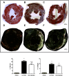
Atorvastatin Improves Ventricular Remodeling after Myocardial Infarction by Interfering with Collagen Metabolism
- PMID: 27880844
- PMCID: PMC5120826
- DOI: 10.1371/journal.pone.0166845
Atorvastatin Improves Ventricular Remodeling after Myocardial Infarction by Interfering with Collagen Metabolism
Erratum in
-
Correction: Atorvastatin Improves Ventricular Remodeling after Myocardial Infarction by Interfering with Collagen Metabolism.PLoS One. 2017 Feb 14;12(2):e0172453. doi: 10.1371/journal.pone.0172453. eCollection 2017. PLoS One. 2017. PMID: 28196119 Free PMC article.
Abstract
Purpose: Therapeutic strategies that modulate ventricular remodeling can be useful after acute myocardial infarction (MI). In particular, statins may exert effects on molecular pathways involved in collagen metabolism. The aim of this study was to determine whether treatment with atorvastatin for 4 weeks would lead to changes in collagen metabolism and ventricular remodeling in a rat model of MI.
Methods: Male Wistar rats were used in this study. MI was induced in rats by ligation of the left anterior descending coronary artery (LAD). Animals were randomized into three groups, according to treatment: sham surgery without LAD ligation (sham group, n = 14), LAD ligation followed by 10mg atorvastatin/kg/day for 4 weeks (atorvastatin group, n = 24), or LAD ligation followed by saline solution for 4 weeks (control group, n = 27). After 4 weeks, hemodynamic characteristics were obtained by a pressure-volume catheter. Hearts were removed, and the left ventricles were subjected to histologic analysis of the extents of fibrosis and collagen deposition, as well as the myocyte cross-sectional area. Expression levels of mediators involved in collagen metabolism and inflammation were also assessed.
Results: End-diastolic volume, fibrotic content, and myocyte cross-sectional area were significantly reduced in the atorvastatin compared to the control group. Atorvastatin modulated expression levels of proteins related to collagen metabolism, including MMP1, TIMP1, COL I, PCPE, and SPARC, in remote infarct regions. Atorvastatin had anti-inflammatory effects, as indicated by lower expression levels of TLR4, IL-1, and NF-kB p50.
Conclusion: Treatment with atorvastatin for 4 weeks was able to attenuate ventricular dysfunction, fibrosis, and left ventricular hypertrophy after MI in rats, perhaps in part through effects on collagen metabolism and inflammation. Atorvastatin may be useful for limiting ventricular remodeling after myocardial ischemic events.
Conflict of interest statement
The authors have declared that no competing interests exist.
Figures


Similar articles
-
Therapeutic effects of continuous infusion of brain natriuretic peptides on postmyocardial infarction ventricular remodelling in rats.Arch Cardiovasc Dis. 2011 Jan;104(1):17-28. doi: 10.1016/j.acvd.2010.09.006. Epub 2010 Dec 17. Arch Cardiovasc Dis. 2011. PMID: 21276574
-
Atorvastatin reduces myocardial fibrosis in a rat model with post-myocardial infarction heart failure by increasing the matrix metalloproteinase-2/tissue matrix metalloproteinase inhibitor-2 ratio.Chin Med J (Engl). 2013;126(11):2149-56. Chin Med J (Engl). 2013. PMID: 23769575
-
Angiotensin type 2 receptor stimulation ameliorates left ventricular fibrosis and dysfunction via regulation of tissue inhibitor of matrix metalloproteinase 1/matrix metalloproteinase 9 axis and transforming growth factor β1 in the rat heart.Hypertension. 2014 Mar;63(3):e60-7. doi: 10.1161/HYPERTENSIONAHA.113.02522. Epub 2013 Dec 30. Hypertension. 2014. PMID: 24379181
-
Remodeling of the myocardium and potential targets in the collagen degradation and synthesis pathways.Curr Drug Targets Cardiovasc Haematol Disord. 2003 Mar;3(1):1-30. doi: 10.2174/1568006033337276. Curr Drug Targets Cardiovasc Haematol Disord. 2003. PMID: 12769643 Review.
-
Ventricular remodeling in heart failure: the role of myocardial collagen.Adv Exp Med Biol. 1995;382:239-45. doi: 10.1007/978-1-4615-1893-8_24. Adv Exp Med Biol. 1995. PMID: 8540401 Review.
Cited by
-
Compound Danshen Tablets ameliorate myocardial ischemia/reperfusion injury-induced ventricular remodeling by regulating autophagy via AMPK/mTOR signaling pathway.Chin Herb Med. 2024 Apr 24;17(3):548-554. doi: 10.1016/j.chmed.2024.03.003. eCollection 2025 Jul. Chin Herb Med. 2024. PMID: 40734915 Free PMC article.
-
Pin1 facilitates isoproterenol‑induced cardiac fibrosis and collagen deposition by promoting oxidative stress and activating the MEK1/2‑ERK1/2 signal transduction pathway in rats.Int J Mol Med. 2018 Mar;41(3):1573-1583. doi: 10.3892/ijmm.2017.3354. Epub 2017 Dec 29. Int J Mol Med. 2018. PMID: 29286102 Free PMC article.
-
Differential effects of Losartan and Atorvastatin in partial and full thickness burn wounds.PLoS One. 2017 Jun 14;12(6):e0179350. doi: 10.1371/journal.pone.0179350. eCollection 2017. PLoS One. 2017. PMID: 28614412 Free PMC article.
-
Traditional Chinese Medicine Intervenes Ventricular Remodeling Following Acute Myocardial Infarction: Evidence From 40 Random Controlled Trials With 3,659 Subjects.Front Pharmacol. 2021 Aug 31;12:707394. doi: 10.3389/fphar.2021.707394. eCollection 2021. Front Pharmacol. 2021. PMID: 34531742 Free PMC article.
-
Polymers and Nanoparticles for Statin Delivery: Current Use and Future Perspectives in Cardiovascular Disease.Polymers (Basel). 2021 Feb 26;13(5):711. doi: 10.3390/polym13050711. Polymers (Basel). 2021. PMID: 33652927 Free PMC article. Review.
References
-
- Sutton MG, Sharpe N. Left ventricular remodeling after myocardial infarction: pathophysiology and therapy. Circulation. 2000;101(25):2981–8. - PubMed
-
- Zornoff LA, Paiva SA, Duarte DR, Spadaro J. Ventricular remodeling after myocardial infarction: concepts and clinical implications. Arquivos brasileiros de cardiologia. 2009;92(2):150–64. - PubMed
-
- Nwogu JI, Geenen D, Bean M, Brenner MC, Huang X, Buttrick PM. Inhibition of collagen synthesis with prolyl 4-hydroxylase inhibitor improves left ventricular function and alters the pattern of left ventricular dilatation after myocardial infarction. Circulation. 2001;104(18):2216–21. - PubMed
-
- Goldsmith EC, Hoffman A, Morales MO, Potts JD, Price RL, McFadden A, et al. Organization of fibroblasts in the heart. Developmental dynamics: an official publication of the American Association of Anatomists. 2004;230(4):787–94. - PubMed
MeSH terms
Substances
LinkOut - more resources
Full Text Sources
Other Literature Sources
Research Materials
Miscellaneous

