. 2016 Nov 22;17(9):2183-2194.
doi: 10.1016/j.celrep.2016.11.005.

RNF166 Determines Recruitment of Adaptor Proteins during Antibacterial Autophagy

Affiliations

RNF166 Determines Recruitment of Adaptor Proteins during Antibacterial Autophagy

Robert J Heath et al. Cell Rep. .

Abstract

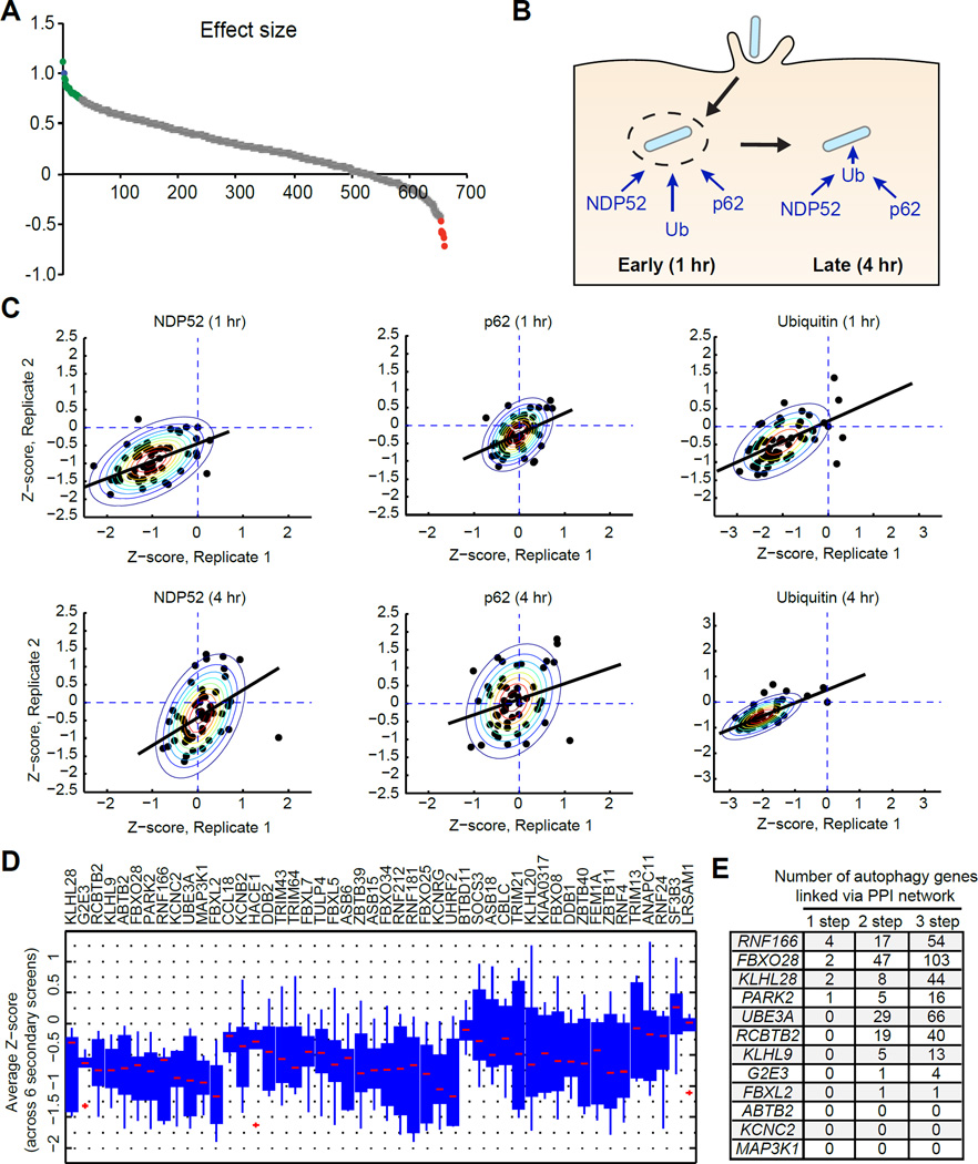
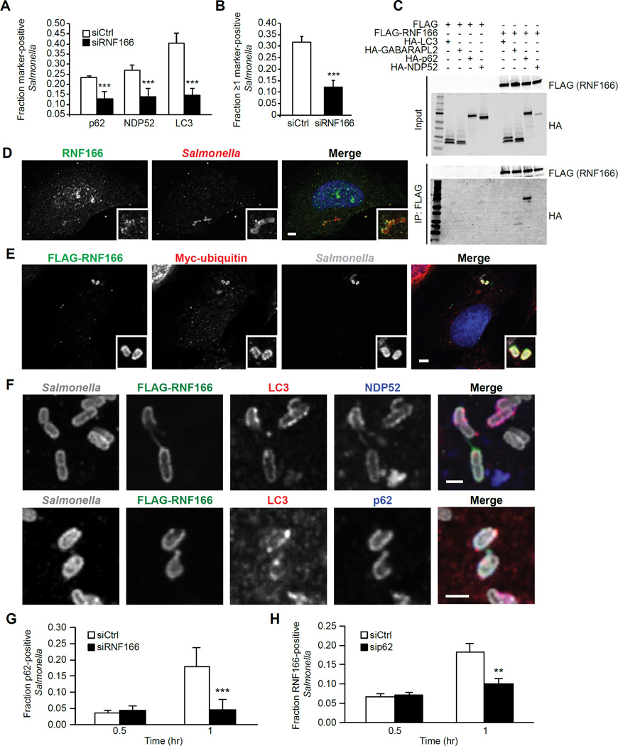
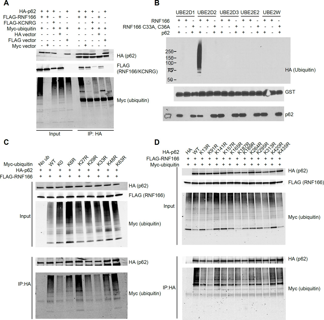
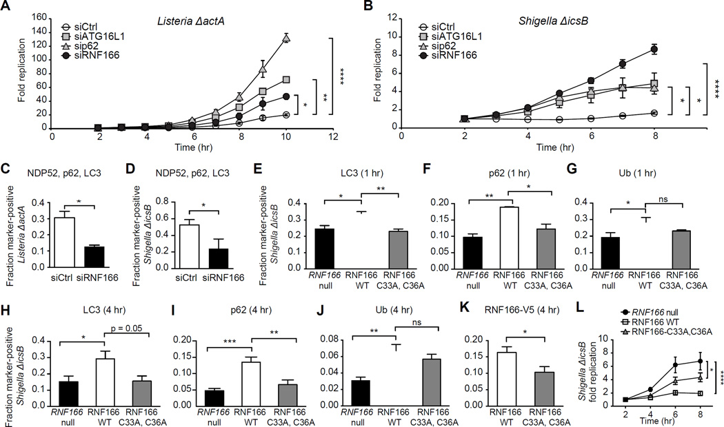
Xenophagy is a form of selective autophagy that involves the targeting and elimination of intracellular pathogens through several recognition, recruitment, and ubiquitination events. E3 ubiquitin ligases control substrate selectivity in the ubiquitination cascade; however, systematic approaches to map the role of E3 ligases in antibacterial autophagy have been lacking. We screened more than 600 putative human E3 ligases, identifying E3 ligases that are required for adaptor protein recruitment and LC3-bacteria colocalization, critical steps in antibacterial autophagy. An unbiased informatics approach pinpointed RNF166 as a key gene that interacts with the autophagy network and controls the recruitment of ubiquitin as well as the autophagy adaptors p62 and NDP52 to bacteria. Mechanistic studies demonstrated that RNF166 catalyzes K29- and K33-linked polyubiquitination of p62 at residues K91 and K189. Thus, our study expands the catalog of E3 ligases that mediate antibacterial autophagy and identifies a critical role for RNF166 in this process.

Keywords: E3 ligases; RNF166; antibacterial autophagy; autophagy; p62; p62 ubiquitination; xenophagy.


Figures

Figure 1
Figure 1. A Core Contingent of E3 Ubiquitin Ligases Function Throughout Antibacterial Autophagy
(A) Distribution of effect size for each siRNA on GFP-LC3-Salmonella colocalization at 1 hr postinfection. Effect size was calculated as a fraction relative to a non-targeting negative control siRNA and a positive control siRNA targeting ATG16L1. Genes that significantly decreased GFP-LC3-Salmonella colocalization are shown in green. Genes that significantly increased GFP-LC3-Salmonella colocalization are shown in red and genes that did not significantly change colocalization are shown in gray. (B) Schematic of antibacterial autophagy targeting strategies. During early bacterial targeting (1 hr), autophagy adaptors can bind independently of ubiquitin. During late bacterial targeting (4 hr), autophagy adaptor proteins bind via ubiquitinated substrates. (C) Z-normalized secondary screen colocalization data. Scatter plots show data from two independent runs for either p62, NDP52, or ubiquitin colocalization at the given time point. Raw data was standardized with z-score computation using mean and standard deviation of negative controls. Contour plots show bivariate Gaussian distribution fit to the data. Black line shows linear regression function fit to the normalized data. (D) Box plots show pooled average Z-scores from each screen per gene. Genes are ordered based on the number of times the average Z-score was negative for all secondary screens and the magnitude of the Z-score. (E) Table of 12 autophagy genes that can be linked to each candidate gene (column 1) via 1-, 2- or 3-step protein-protein interactions derived from Bioplex database. See also Figure S1, Tables S1–S2.
Figure 2
Figure 2. RNF166 Is Required to Recruit the Autophagy Apparatus to Salmonella and Interacts with p62
(A) HeLa cells were treated with a non-targeting siRNA or siRNA targeting RNF166 for 48 hr. Cells were then infected with Salmonella for 1 hr and the fraction of Salmonella colocalizing with either p62, NDP52, or LC3 was enumerated. Data represent means ± SEM, n = 100 infected cells per group, data pooled from three independent experiments. (B) RNF166-depleted HeLa cells were infected with Salmonella for 1 hr and co-stained for endogenous LC3, p62, and NDP52. The fraction of all intracellular bacteria that colocalized with ≥ 1 marker (LC3, p62, and/or NDP52) was determined. Data represent means ± SEM, n = 100 infected cells per group, data pooled from three independent experiments. (C) HEK293T cells were transfected for 24 hr with constructs expressing FLAG alone or FLAGRNF166 and HA-tagged autophagy proteins as indicated. Proteins were immunoprecipitated with anti-FLAG antibodies. Data are representative of four independent experiments. (D) Confocal images of HeLa cells infected with Salmonella for 1 hr and stained for endogenous RNF166. Insets indicate areas of bacterial colocalization. Data are representative of three independent experiments. (E) Confocal images of HeLa cells transfected for 24 hr with FLAG-tagged RNF166 and Myctagged ubiquitin infected with Salmonella for 1 hr. Insets indicate areas of bacterial colocalization with FLAG-RNF166 and Myc-ubiquitin. Data are representative of three independent experiments. (F) Confocal images of HeLa cells transfected for 24 hr with FLAG-tagged RNF166 and infected with Salmonella for 1 hr. Cells expressing FLAG-RNF166 were co-stained for LC3 and NDP52 (top panels) or LC3 and p62 (bottom panels). Data are representative of three independent experiments. (G) HeLa cells treated with a non-targeting siRNA or siRNA targeting RNF166 for 48 hr were infected with Salmonella for the indicated time periods. Cells were stained for endogenous p62. Shown is the fraction of Salmonella colocalizing with p62 enumerated for at least 100 cells. Data represent means ± SEM, n = 100 infected cells per group, data pooled from three independent experiments. (H) HeLa cells were treated with a non-targeting siRNA or siRNA targeting p62 for 48 hr, infected with Salmonella for the indicated time periods, and stained for endogenous RNF166. Shown is the fraction of Salmonella colocalizing with RNF166 at the indicated time points. Data represent means ± SEM, n = 150 infected cells per group, data pooled from three independent experiments. For all panels, **p < 0.01, ***p < 0.001, Student’s t test. See also Figure S2.
Figure 3
Figure 3. RNF166 Mediates K29- and K33-Linked Ubiquitination of p62
(A) HEK293T cells were transfected with indicated constructs followed by immunoprecipitation of HA-p62. Representative blot is shown from four independent experiments. (B) GST-RNF166 and SUMO-p62 were incubated together or separately in the presence of recombinant UBE1 (E1), various E2 ubiquitin-conjugating enzymes as indicated, and HAubiquitin. Representative blot is shown from three independent experiments. (C) HEK293T cells were transfected with FLAG-RNF166, HA-p62 and one of the indicated Mycubiquitin constructs with single point mutations at the indicated lysine residues. Proteins were immunoprecipitated with anti-HA antibodies and immunoblots were performed with antibodies against HA and Myc to detect ubiquitinated proteins. Representative blot is shown from four independent experiments. (D) HEK293T cells were co-transfected with Myc-ubiquitin, FLAG-RNF166, and HA-p62 with the indicated mutations. Proteins were immunoprecipitated with anti-HA antibodies and immunoblots were performed with antibodies against HA and Myc to detect ubiquitinated proteins. Representative blot is shown from three independent experiments.
Figure 4
Figure 4. RNF166 Is Required to Inhibit the Intracellular Replication of Listeria and Shigella
(A) HeLa cells treated with a non-targeting siRNA or siRNA targeting RNF166, p62, or ATG16L1 for 48 hr were infected with Listeria ΔactA expressing luciferase. Cells were treated with gentamicin to remove extracellular bacteria and relative light units were monitored over the indicated time course. Fold replication represents light units over time compared to 2 hr postinfection. Data represent means ± SEM, n = 8. (B) Cells were treated as in (A) and infected with Shigella ΔicsB expressing luciferase. Data represent means ± SEM, n = 8. (C, D) HeLa cells were treated with a non-targeting siRNA or siRNA targeting RNF166 for 48 hr, then infected with Listeria ΔactA (C) or Shigella ΔicsB (D) for 1 hr and co-stained for endogenous NDP52, p62, and LC3. The fraction of colocalization of each intracellular bacterium simultaneously with NDP52, p62, and LC3 was scored. > 50 bacteria from three independent experiments were analyzed. Data represent means + SEM. (E-G) Quantification of LC3 (E), p62 (F), and ubiquitin (G) recruitment to Shigella ΔicsB at 1 hr postinfection in RNF166-null HeLa cells expressing the indicated constructs. Data represent means ± SEM, n = 125 infected cells per group, data pooled from three independent experiments. (H-K) Quantification of LC3 (H), p62 (I), ubiquitin (J), and RNF166-V5 (K) recruitment to Shigella ΔicsB in RNF166-null HeLa expressing the indicated constructs. Data represent means ± SEM, n = 125 infected cells per group, data pooled from three independent experiments. (L) Intracellular replication of Shigella ΔicsB expressing luciferase in RNF166-null HeLa cells expressing the indicated constructs. Cells were treated with gentamicin to remove extracellular bacteria and relative light units were monitored over the indicated time course. Fold replication represents light units over time compared to 2 hr postinfection. Data represent means ± SEM, n = 8. For all panels, *p < 0.05; **p < 0.01; ***p < 0.001; ****p < 0.0001; ns, not significant. Student’s t test for (C, D, K), one-way ANOVA with multiple comparisons for (A, B, E-J, and L). See also Figures S3 and S4.

References

    1. Antonioli M, Albiero F, Nazio F, Vescovo T, Perdomo AB, Corazzari M, Marsella C, Piselli P, Gretzmeier C, Dengjel J, et al. AMBRA1 interplay with cullin E3 ubiquitin ligases regulates autophagy dynamics. Dev. Cell. 2014;31:734–746. - PubMed
    1. Begun J, Lassen KG, Jijon HB, Baxt LA, Goel G, Heath RJ, Ng A, Tam JM, Kuo SY, Villablanca EJ, et al. Integrated Genomics of Crohn's Disease Risk Variant Identifies a Role for CLEC12A in Antibacterial Autophagy. Cell Rep. 2015;11:1905–1918. - PMC - PubMed
    1. Birmingham CL, Smith AC, Bakowski MA, Yoshimori T, Brumell JH. Autophagy controls Salmonella infection in response to damage to the Salmonella-containing vacuole. J. Biol. Chem. 2006;281:11374–11383. - PubMed
    1. Case ED, Chong A, Wehrly TD, Hansen B, Child R, Hwang S, Virgin HW, Celli J. The Francisella O-antigen mediates survival in the macrophage cytosol via autophagy avoidance. Cell. Microbiol. 2014;16:862–877. - PMC - PubMed
    1. Cemma M, Kim PK, Brumell JH. The ubiquitin-binding adaptor proteins p62/SQSTM1 and NDP52 are recruited independently to bacteria-associated microdomains to target Salmonella to the autophagy pathway. Autophagy. 2011;7:341–345. - PMC - PubMed

MeSH terms