Review
. 2017 Jan;97(1):227-252.
doi: 10.1152/physrev.00037.2015.

Cardiac T-Tubule Microanatomy and Function

Affiliations
Review

Cardiac T-Tubule Microanatomy and Function

TingTing Hong et al. Physiol Rev. 2017 Jan.

Abstract

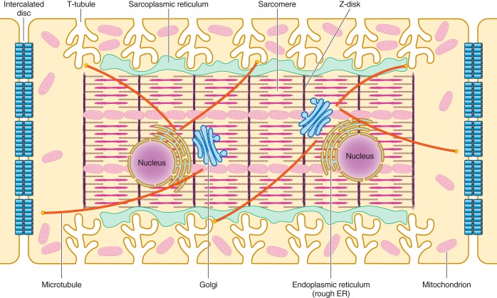
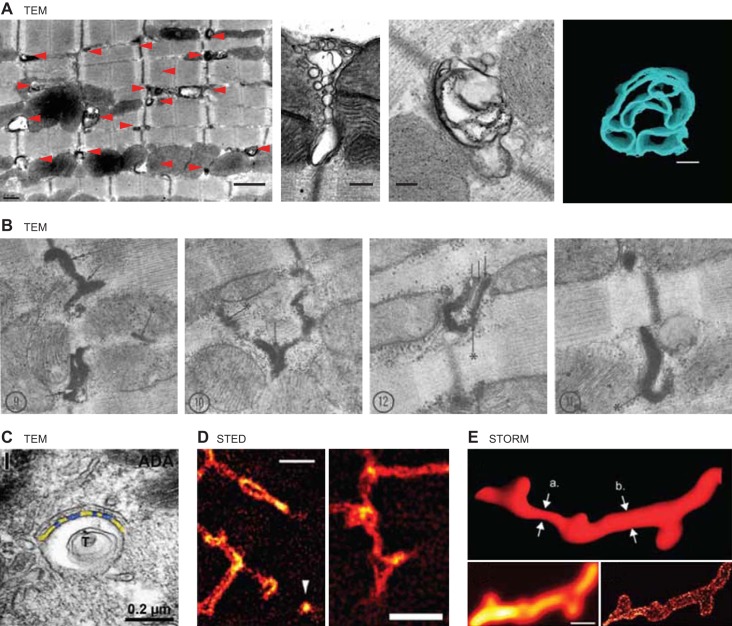
Unique to striated muscle cells, transverse tubules (t-tubules) are membrane organelles that consist of sarcolemma penetrating into the myocyte interior, forming a highly branched and interconnected network. Mature t-tubule networks are found in mammalian ventricular cardiomyocytes, with the transverse components of t-tubules occurring near sarcomeric z-discs. Cardiac t-tubules contain membrane microdomains enriched with ion channels and signaling molecules. The microdomains serve as key signaling hubs in regulation of cardiomyocyte function. Dyad microdomains formed at the junctional contact between t-tubule membrane and neighboring sarcoplasmic reticulum are critical in calcium signaling and excitation-contraction coupling necessary for beat-to-beat heart contraction. In this review, we provide an overview of the current knowledge in gross morphology and structure, membrane and protein composition, and function of the cardiac t-tubule network. We also review in detail current knowledge on the formation of functional membrane subdomains within t-tubules, with a particular focus on the cardiac dyad microdomain. Lastly, we discuss the dynamic nature of t-tubules including membrane turnover, trafficking of transmembrane proteins, and the life cycles of membrane subdomains such as the cardiac BIN1-microdomain, as well as t-tubule remodeling and alteration in diseased hearts. Understanding cardiac t-tubule biology in normal and failing hearts is providing novel diagnostic and therapeutic opportunities to better treat patients with failing hearts.


Figures

FIGURE 1.
FIGURE 1.
Schematic illustration of the internal structures of an adult ventricular cardiomyocyte. T-tubules, which are enriched with voltage-gated L-type calcium channels, are positioned closely near the sarcoplasmic reticulum, the primary internal calcium store. Sarcomeres form myofibrils, which are responsible for cardiomyocyte contraction upon calcium release. The Golgi apparatus and microtubules serve as the “loading dock” and “highways,” respectively, to deliver ion channels to specific subdomains on the plasma membrane. Mitochondria provide the energy needed for the contraction of cardiomyocytes. Intercalated discs located at the longitudinal sides of each ventricular cardiomyocyte mediate the cell-to-cell propagation of action potentials.
FIGURE 2.
FIGURE 2.
Images of t-tubule microfolds. A: TEM images reproduced from Hong et al. (91). B: TEM images reproduced from Forssmann and Girardier (62). C: TEM image reproduced from Lavorato et al. (117), with permission from Springer. D: STED images reproduced from Wagner et al. (220). E: STORM images reproduced from Jayasinghe et al. (101).
FIGURE 3.
FIGURE 3.
Bin1 splice variants in adult mouse cardiomyocytes. Cartoon of Bin1 exons and the splice variants found in adult mouse cardiomyocytes, as well as brain and skeletal muscle. BAR, Bin-amphiphysin-Rvs domain; PI, phosphoinositide binding domain; CLAP, clathrin/AP2 binding region; MBD, myc binding domain; SH3, SH3 domain. For details, see Reference 89.
FIGURE 4.
FIGURE 4.
Schematic illustration of BIN1-microdomain organization within cardiac t-tubules. Top: the “banana”-shaped molecule cBIN1 regulates cardiac t-tubule function through: 1) facilitating microtubule-dependent forward trafficking of Cav1.2 channels (LTCC, L-type calcium channels) to cBIN1 organized membrane microfolds at t-tubules (targeted delivery); 2) clustering of LTCCs and RyRs at cBIN1-microfolds based microdomains; 3) creating extracellular ion slow diffusion zone within t-tubule lumen; 4) organizing microdomains for dyad formation and regulation by β-adrenergic signaling. Bottom: cartoon of mammalian adult ventricular cardiomyocytes, which are rod-shaped striated muscle cells with t-tubule invaginations occurring periodically near z-discs of myofilaments.
FIGURE 5.
FIGURE 5.
Schematic representation of ion channel targeted delivery. Channel proteins are sorted into vesicular carriers and docked onto microtubules at the trans-Golgi network (TGN) and subsequently delivered to their subcellular destinations in cooperation with actin “rest stops” along the route. Microtubule plus-end binding proteins interact with anchor proteins of specific membrane subdomains, allowing targeted delivery of cargo proteins. In the case of connexin 43 (Cx43) trafficking, the interaction between the microtubule plus-end binding protein EB1, the channel itself, and the adherens junction complex ensures the targeted delivery of Cx43 hemichannels to the intercalated discs. For LTCC delivery to t-tubules, key components are the LTCC channels, a +TIP protein, and cardiac bridging integrator 1 (cBIN1). The microtubule +TIP protein associated with LTCC delivery has not yet been identified.
FIGURE 6.
FIGURE 6.
Alternative translation of connexin 43. In addition to full-length 43-kDa connexin 43 (GJA1-43K), there are six NH2-terminal truncated smaller Cx43 isoforms (GJA1-32K, GJA1-29K, GJA1-26K, GJA1-20K, GJA1-11K, and GJA1-7K) resulting from internal methionine residues that initiate ribosomal translation, a process known as alternative translation. At least one of the isoform, GJA1-20k, has been identified as necessary for the full-length channel to traffick to the surface membrane.
FIGURE 7.
FIGURE 7.
Cartoon of BIN1-dependent recruitment of phosphorylated ryanodine receptors (RyR) to dyads during acute stress. Acute isoproterenol (ISO) redistributes BIN1 to cardiac t-tubules and subsequently recruits phosphorylated RyRs to couple with LTCCs at dyads, increasing excitation-contraction coupling gain while maintaining electrical stability. In Bin1 HT cardiomyocytes with less BIN1 available, insufficient RyR recruitment leads to accumulation of hyperactive phosphorylated RyRs outside of dyads, increasing spontaneous calcium release and promoting arrhythmias.
FIGURE 8.
FIGURE 8.
Schematic of endocytosis from cBIN1 microdomains. Adapting BAR domain regulated endocytosis from noncardiac cells (229), we expect that for t-tubules, cBIN1 interacts with cortical actin to generate an endocytic neck, and the SH3 domain of BIN1 the binds to dynamin 2 for scission and internalization of the endocytic vesicle.
FIGURE 9.
FIGURE 9.
BIN1-microdomain release from cardiac t-tubules. A schematic of how fission can potentially occur between two neighboring invaginated cBIN1 microfolds, resulting in external release of cBIN1 microfolds that result in blood-borne microparticles.
FIGURE 10.
FIGURE 10.
Reduced cBIN1 microdomains in failing cardiomyocytes. In failing cardiomyocytes with reduced cBIN1 and cBIN1-induced microdomains, expression of ion channels on the cell surface and the morphology of t-tubules are altered. As highlighted in the cartoon, changes following reduced BIN1 transcription include 1) impaired microtubule-dependent targeted delivery of LTCC results in reduced t-tubule surface expression of LTCCs; 2) disruption of cBIN1 microdomains impairs sufficient LTCC clustering within t-tubules; 3) loss of dense membrane microfolds and resultant t-tubule dilation cause removal of the protective slow diffusion zone within t-tubule lumen; and 4) impaired recruitment of RyRs causes accumulation of hyperphosphorylated RyRs outside dyads, increasing orphaned leaky RyRs.

References

    1. Arakel EC, Brandenburg S, Uchida K, Zhang H, Lin YW, Kohl T, Schrul B, Sulkin MS, Efimov IR, Nichols CG, Lehnart SE, Schwappach B. Tuning the electrical properties of the heart by differential trafficking of KATP ion channel complexes. J Cell Sci : 2106–2119, 2014. - PMC - PubMed
    1. Asghari P, Scriven DR, Sanatani S, Gandhi SK, Campbell AI, Moore ED. Nonuniform and variable arrangements of ryanodine receptors within mammalian ventricular couplons. Circ Res : 252–262, 2014. - PubMed
    1. Ayettey AS, Navaratnam V. The T-tubule system in the specialized and general myocardium of the rat. J Anat : 125–140, 1978. - PMC - PubMed
    1. Balijepalli RC, Foell JD, Hall DD, Hell JW, Kamp TJ. Localization of cardiac L-type Ca2+ channels to a caveolar macromolecular signaling complex is required for beta(2)-adrenergic regulation. Proc Natl Acad Sci USA : 7500–7505, 2006. - PMC - PubMed
    1. Balijepalli RC, Kamp TJ. Caveolae, ion channels and cardiac arrhythmias. Prog Biophys Mol Biol : 149–160, 2008. - PMC - PubMed

Publication types

LinkOut - more resources