. 2017 Jan 18;91(3):e01656-16.
doi: 10.1128/JVI.01656-16. Print 2017 Feb 1.

Activation of COX-2/PGE2 Promotes Sapovirus Replication via the Inhibition of Nitric Oxide Production

Affiliations

Activation of COX-2/PGE2 Promotes Sapovirus Replication via the Inhibition of Nitric Oxide Production

Mia Madel Alfajaro et al. J Virol. .

Abstract

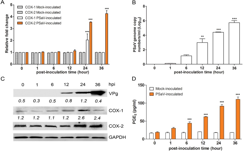
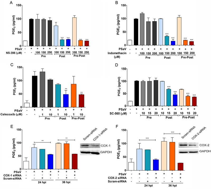
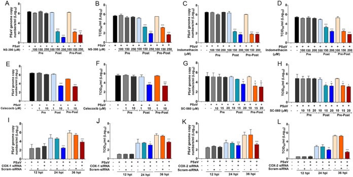
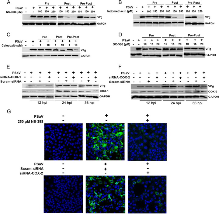
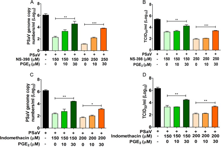
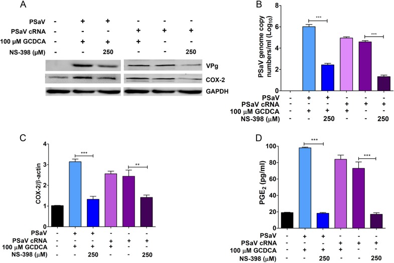
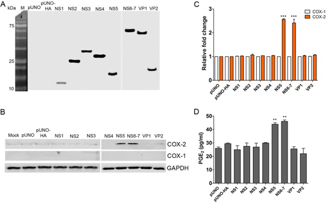
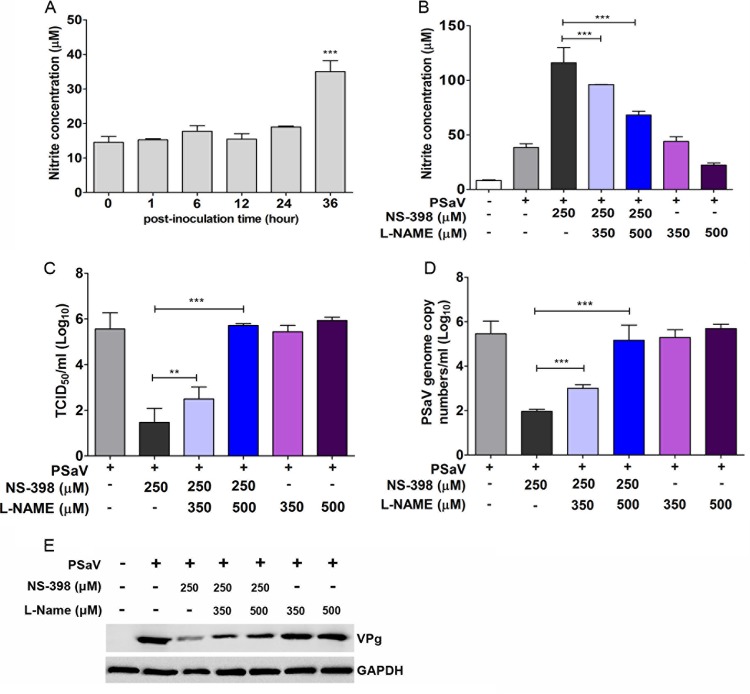
Enteric caliciviruses in the genera Norovirus and Sapovirus are important pathogens that cause severe acute gastroenteritis in both humans and animals. Cyclooxygenases (COXs) and their final product, prostaglandin E2 (PGE2), are known to play important roles in the modulation of both the host response to infection and the replicative cycles of several viruses. However, the precise mechanism(s) by which the COX/PGE2 pathway regulates sapovirus replication remains largely unknown. In this study, infection with porcine sapovirus (PSaV) strain Cowden, the only cultivable virus within the genus Sapovirus, markedly increased COX-2 mRNA and protein levels at 24 and 36 h postinfection (hpi), with only a transient increase in COX-1 levels seen at 24 hpi. The treatment of cells with pharmacological inhibitors, such as nonsteroidal anti-inflammatory drugs or small interfering RNAs (siRNAs) against COX-1 and COX-2, significantly reduced PGE2 production, as well as PSaV replication. Expression of the viral proteins VPg and ProPol was associated with activation of the COX/PGE2 pathway. We observed that pharmacological inhibition of COX-2 dramatically increased NO production, causing a reduction in PSaV replication that could be restored by inhibition of nitric oxide synthase via the inhibitor N-nitro-l-methyl-arginine ester. This study identified a pivotal role for the COX/PGE2 pathway in the regulation of NO production during the sapovirus life cycle, providing new insights into the life cycle of this poorly characterized family of viruses. Our findings also reveal potential new targets for treatment of sapovirus infection.

Importance: Sapoviruses are among the major etiological agents of acute gastroenteritis in both humans and animals, but little is known about sapovirus host factor requirements. Here, using only cultivable porcine sapovirus (PSaV) strain Cowden, we demonstrate that PSaV induced the vitalization of the cyclooxygenase (COX) and prostaglandin E2 (PGE2) pathway. Targeting of COX-1/2 using nonsteroidal anti-inflammatory drugs (NSAIDs) such as the COX-1/2 inhibitor indomethacin and the COX-2-specific inhibitors NS-398 and celecoxib or siRNAs targeting COXs, inhibited PSaV replication. Expression of the viral proteins VPg and ProPol was associated with activation of the COX/PGE2 pathway. We further demonstrate that the production of PGE2 provides a protective effect against the antiviral effector mechanism of nitric oxide. Our findings uncover a new mechanism by which PSaV manipulates the host cell to provide an environment suitable for efficient viral growth, which in turn can be a new target for treatment of sapovirus infection.

Keywords: caliciviruses; cyclooxygenases; nitric oxide; prostaglandin E2; sapovirus.


Figures

FIG 1
FIG 1
Induction of COX-1 and COX-2 by PSaV infection. (A and B) The expression of COX-1, COX-2, and PSaV viral RNA in LLC-PK cells infected with PSaV (MOI = 1 FFU/cell) was quantified by real-time PCR (qPCR). In the cases of COX-1 and COX-2, expression levels were normalized to β-actin and are depicted as the fold induction compared with that of the mock-inoculated cells. (C) The levels of the VPg, COX-1, COX-2, and GAPDH proteins were analyzed by Western blotting. GAPDH was used as a loading control. (D) The levels of PGE2 in the supernatants harvested at 36 hpi from PSaV-infected LLC-PK cells were determined by ELISA. The levels of PGE2 in the supernatants were compared between mock- and virus-inoculated groups. The data are presented as means and standard errors of the mean from the results of three independent experiments. Differences were evaluated using one-way analysis of variance (ANOVA). **, P < 0.001; ***, P < 0.0001.
FIG 2
FIG 2
Effects of COX-2 inhibitors on PGE2 production during PSaV infection. (A to D) LLC-PK cells were treated with selective COX-2 inhibitors (NS398 and celecoxib), a nonselective COX inhibitor (indomethacin), and a selective COX-1 inhibitor (SC-560) as indicated prior to the addition of the virus inoculum (MOI = 1 FFU/cell), and then the inhibitor(s) was removed (Pre); after the addition of the virus inoculum, and then the inhibitor(s) was left for the duration of the infection (Post); or prior to the addition of the inoculum, as well as for the duration of the infection (Pre-Post). The levels of PGE2 in the supernatants harvested at 36 hpi were determined by ELISA. The levels of PGE2 in the supernatants of virus-infected cultures were compared between the mock- and chemical-treated groups. (E and F) Confluent LLC-PK cells were transfected with siRNAs against COX-1, COX-2, or scrambled siRNA (Scram-siRNA) prior to infection with PSaV (MOI = 1 FFU/cell). The supernatants were collected, and ELISA was conducted to determine the PGE2 concentrations. The levels of PGE2 in the supernatants were compared between mock- and siRNA-transfected groups. (Insets) Western blot analysis for COX-1, COX-2, and GAPDH was conducted with LLC-PK cells transfected with COX-1, COX-2, or scrambled siRNA. The data are presented as means and standard errors of the mean from the results of three independent experiments. Differences were evaluated using one-way ANOVA. **, P < 0.001; ***, P < 0.0001.
FIG 3
FIG 3
Inhibition of COXs attenuates PSaV replication. (A to H) LLC-PK cells were pretreated (Pre), posttreated (Post), or pre/posttreated (Pre-Post) with noncytotoxic doses of NS-398, indomethacin, celecoxib, and SC560. At 36 hpi with PSaV (MOI = 1 FFU/cell), cells were harvested, and the levels of viral RNA (A, C, E, and G) and the titer (B, D, F, and H) were determined by quantitative real-time PCR and TCID50, respectively. (I to L) LLC-PK cells were transfected with siRNAs against COX-1, COX-2, or scrambled siRNA before inoculation with PSaV (MOI = 1 FFU/cell). Samples were harvested at 36 hpi, and the levels of viral RNA (I and K) and the titer (J and L) were determined by quantitative real-time PCR and TCID50, respectively. The data are displayed as means and standard errors of the mean from the results of three independent experiments. Differences were evaluated using one-way ANOVA. *, P < 0.05; **, P < 0.001; ***, P < 0.0001.
FIG 4
FIG 4
Effects of COX inhibitors or siRNA on PSaV replication. (A to D) LLC-PK cells were pretreated (Pre), posttreated (Post), or pre/posttreated (Pre-Post) with noncytotoxic doses of NS-398, indomethacin, celecoxib, and SC560. At 36 hpi with PSaV (MOI = 1 FFU/cell), cells were harvested, and the levels of viral VPg were determined by Western blot analyses. (E and F) LLC-PK cells were transfected with siRNAs against COX-1, COX-2, or scrambled siRNA before inoculation with PSaV (MOI = 1 FFU/cell). Samples were harvested and processed as described above. GAPDH was used as a loading control. (G) LLC-PK cells were infected with PSaV (MOI = 1 FFU/cell), and the effect of the COX-2 inhibitor NS-398 on viral antigen production was determined by confocal microscopy.
FIG 5
FIG 5
Addition of exogenous PGE2 reverses the effects of COX inhibitors on PSaV replication. LLC-PK cells were infected with PSaV (MOI = 1 FFU/cell), treated with noncytotoxic doses of NS-398 or indomethacin, and then supplemented with exogenous PGE2 in the maintenance medium. After 36 h postinfection, cells were harvested, and the levels of viral RNA synthesis (A and C) and the titer (B and D) were determined by quantitative real-time PCR and TCID50, respectively. The data are represented as means and standard errors of the mean from the results of three independent experiments. Differences were evaluated using one-way ANOVA. *, P < 0.05; **, P < 0.001; ***, P < 0.001.
FIG 6
FIG 6
Bile acid GCDCA does not influence the expression of COX-2 during PSaV infection. (A to C) LLC-PK cells were either infected with PSaV (MOI = 1 FFU/cell) or transfected with 1 μg of in vitro-transcribed PSaV-capped RNA, and the effect of the COX-2 inhibitor NS-398 or the bile acid GCDCA was examined. Infected cells were harvested at 36 h postinfection, whereas transfected cells were harvested after 6 days posttransfection and subjected to Western blot analysis to assess viral protein production (A), as well as to qPCR for viral RNA (B) and COX-2 (C). (D) Supernatants were also collected for ELISA to quantify the levels of PGE2. The data are represented as means and standard errors of the mean from the results of three independent experiments. Differences were evaluated using one-way ANOVA. **, P < 0.001; ***, P < 0.001.
FIG 7
FIG 7
Role of PSaV proteins in stimulating COX-2 expression. (A and B) LLC-PK cells were transfected with 1 μg of pUNO plasmids containing each PSaV gene tagged with an HA epitope, as indicated in Materials and Methods. As controls, pUNO empty or HA-carrying (pUNO-HA) plasmids were transfected. At 36 h posttransfection (hpt), cells were harvested, and the expression levels of viral proteins (A), as well as COX-1 and COX-2 proteins (B), were determined by Western blotting. GAPDH was used as a loading control. (C) In the cases of COX-1 and COX-2, the expression levels were also quantified by real-time PCR and normalized to β-actin and are depicted as the fold induction compared with that of vehicle-transfected cells. (D) To determine the PGE2 concentration, supernatants were collected and ELISA was conducted. The levels of PGE2 in the supernatants were compared between vehicle- and PSaV gene-transfected groups. The data are presented as means and standard errors of the mean from three independent experiments. Differences were evaluated using one-way ANOVA. **, P < 0.001; ***, P < 0.0001.
FIG 8
FIG 8
PGE2 blocks the antiviral effect of nitric oxide on PSaV infection. (A) Supernatants from mock- or PSaV-infected samples were collected, and the nitrite concentration was determined using the Griess reagent system, as described in Materials and Methods. LLC-PK cells were infected with PSaV (MOI = 1 FFU/cell) and subsequently treated with the COX-2 inhibitor NS-398 or the nitric oxide synthase inhibitor L-NAME, either singly or in combination. (B) The effect of inhibitor treatment on nitric oxide production was then determined as described for panel A. (C to E) The levels of viral titer (C), RNA (D), and protein (E) were determined by TCID50, real-time reverse transcription (RT)-PCR, and Western blot analyses, respectively. GAPDH served as the loading control. The data are means and standard errors of the mean from the results of three different independent experiments. Differences were evaluated by one-way ANOVA. **, P < 0.001; ***, P < 0.0001.

References

    1. Anderson EJ. 2010. Prevention and treatment of viral diarrhea in pediatrics. Expert Rev Anti Infect Ther 8:205–217. doi:10.1586/eri.10.1. - DOI - PubMed
    1. Ayukekbong JA, Mesumbe HN, Oyero OG, Lindh M, Berdström T. 2015. Role of noroviruses as aetiological of diarrhea in developing countries. J Gen Virol 96:1983–1999. doi:10.1099/vir.0.000194. - DOI - PubMed
    1. Patel MM, Hall AJ, Vinjé J, Parashar UD. 2009. Noroviruses: a comprehensive review. J Clin Virol 44:1–8. doi:10.1016/j.jcv.2008.10.009. - DOI - PubMed
    1. Jones MK, Watanabe M, Zhu S, Graves CL, Keyes LR, Grau KR, Gonzalez-Hernandez MB, Iovine NM, Wobus CE, Vinjé J, Tibbetts SA, Wallet SM, Karst SM. 2014. Enteric bacteria promote human and mouse norovirus infection of B cells. Science 346:755–759. doi:10.1126/science.1257147. - DOI - PMC - PubMed
    1. Oka T, Wang Q, Katamayan K, Saif LJ. 2015. Comprehensive review of human sapoviruses. Clin Microbiol Rev 28:32–53. doi:10.1128/CMR.00011-14. - DOI - PMC - PubMed

MeSH terms