. 2017 Jan 18;91(3):e02230-16.
doi: 10.1128/JVI.02230-16. Print 2017 Feb 1.

Mechanism of Enhanced HIV Restriction by Virion Coencapsidated Cytidine Deaminases APOBEC3F and APOBEC3G

Affiliations

Mechanism of Enhanced HIV Restriction by Virion Coencapsidated Cytidine Deaminases APOBEC3F and APOBEC3G

Anjuman Ara et al. J Virol. .

Erratum in

Abstract

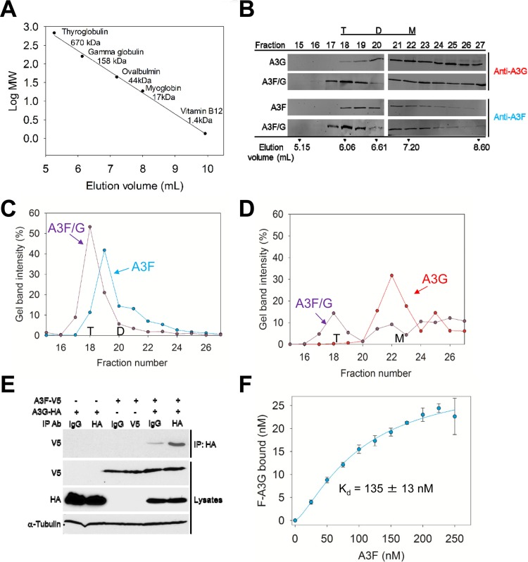
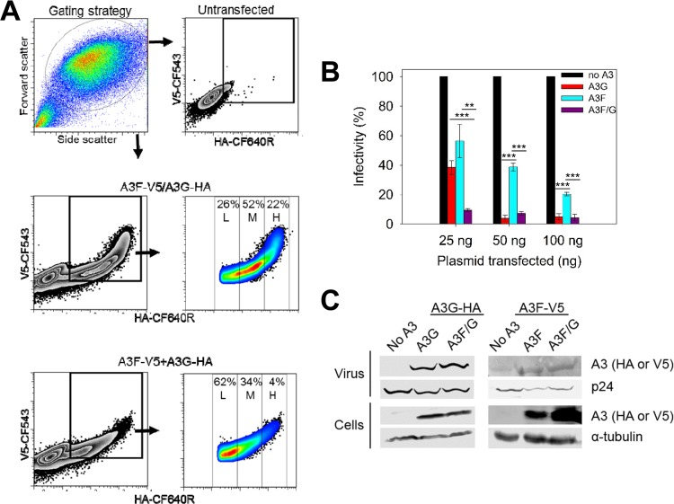
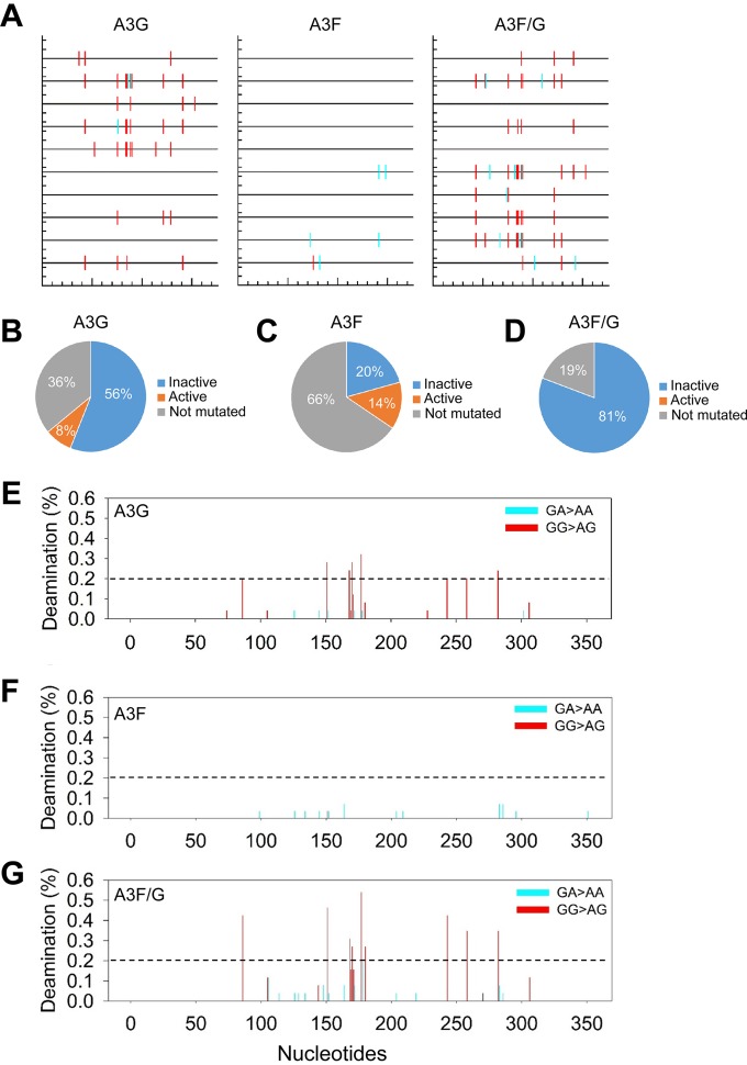
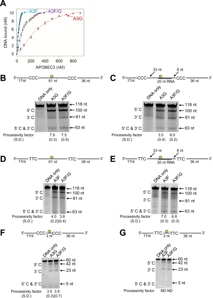
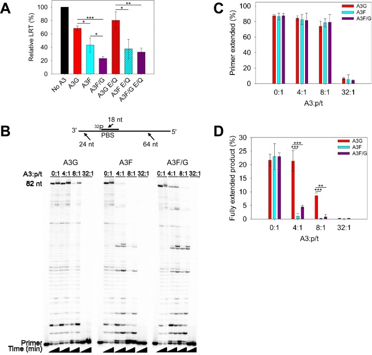
The APOBEC3 (A3) enzymes, A3G and A3F, are coordinately expressed in CD4+ T cells and can become coencapsidated into HIV-1 virions, primarily in the absence of the viral infectivity factor (Vif). A3F and A3G are deoxycytidine deaminases that inhibit HIV-1 replication by inducing guanine-to-adenine hypermutation through deamination of cytosine to form uracil in minus-strand DNA. The effect of the simultaneous presence of both A3G and A3F on HIV-1 restriction ability is not clear. Here, we used a single-cycle infectivity assay and biochemical analyses to determine if coencapsidated A3G and A3F differ in their restriction capacity from A3G or A3F alone. Proviral DNA sequencing demonstrated that compared to each A3 enzyme alone, A3G and A3F, when combined, had a coordinate effect on hypermutation. Using size exclusion chromatography, rotational anisotropy, and in vitro deamination assays, we demonstrate that A3F promotes A3G deamination activity by forming an A3F/G hetero-oligomer in the absence of RNA which is more efficient at deaminating cytosines. Further, A3F caused the accumulation of shorter reverse transcripts due to decreasing reverse transcriptase efficiency, which would leave single-stranded minus-strand DNA exposed for longer periods of time, enabling more deamination events to occur. Although A3G and A3F are known to function alongside each other, these data provide evidence for an A3F/G hetero-oligomeric A3 with unique properties compared to each individual counterpart.

Importance: The APOBEC3 enzymes APOBEC3F and APOBEC3G act as a barrier to HIV-1 replication in the absence of the HIV-1 Vif protein. After APOBEC3 enzymes are encapsidated into virions, they deaminate cytosines in minus-strand DNA, which forms promutagenic uracils that induce transition mutations or proviral DNA degradation. Even in the presence of Vif, footprints of APOBEC3-catalyzed deaminations are found, demonstrating that APOBEC3s still have discernible activity against HIV-1 in infected individuals. We undertook a study to better understand the activity of coexpressed APOBEC3F and APOBEC3G. The data demonstrate that an APOBEC3F/APOBEC3G hetero-oligomer can form that has unique properties compared to each APOBEC3 alone. This hetero-oligomer has increased efficiency of virus hypermutation, raising the idea that we still may not fully realize the antiviral mechanisms of endogenous APOBEC3 enzymes. Hetero-oligomerization may be a mechanism to increase their antiviral activity in the presence of Vif.

Keywords: APOBEC3; DNA-protein interactions; HIV; deaminase; mutagenesis; oligomerization; processivity; protein-protein interactions; reverse transcriptase.


Figures

FIG 1
FIG 1
A3F and A3G hetero-oligomerize. (A to D) Size exclusion chromatography was conducted with a 10-ml G200 Superdex column. (A) A calibration curve was used to calculate the molecular weights (MW) and oligomerization states of the enzymes. (B) The A3G, A3F, and A3F/G SEC experiments used quantitative immunoblotting to detect A3G or A3F with antibodies to the native proteins. The integrated band intensities calculated using LiCor/Odyssey software were used to generate chromatograms. The T, D, and M notations indicate peak fractions for tetramers, dimers, and monomers, respectively. (C) The integrated band intensities for A3F indicated that A3F alone was primarily a trimer (fraction 19, 153 kDa) with a minority of dimers (fraction 20, 101 kDa). The A3F in the A3F/G combined run was primarily a tetramer (fraction 18, 207 kDa). (D) The integrated band intensities for A3G indicated that A3G alone was primarily a monomer (fraction 22, 46 kDa) with a minority of dimers (fraction 20, 101 kDa). The A3G in the A3F/G combined run maintained a population of monomers (fraction 22) but also was able to fractionate with the peak fraction corresponding to tetramers (fraction 18, 207 kDa). (E) Coimmunoprecipitation of A3F-V5 with A3G-HA. A3G-HA and A3F-V5 were transfected alone or in combination. The immunoprecipitation of cell lysates used either anti-HA antibody or rabbit IgG (mock), and samples were immunoblotted with antibodies against α-tubulin, HA, and V5. Cell lysates show the expression of α-tubulin, HA, and V5. (F) Steady-state fluorescence depolarization was used to measure the rotational anisotropy of F-A3G interacting with A3F. Rotational anisotropy was normalized to fraction F-A3G bound. An apparent Kd was calculated by regression analysis of the saturation curve from three independent experiments. A sigmoidal fit was chosen by least-squares analysis and resulted in an apparent Kd of 135 ± 13 nM.
FIG 2
FIG 2
Coexpressed A3F and A3G enhances the restriction of HIV replication. (A) Flow cytometry was used to detect A3G-HA or A3F-V5 in individual cells after transient transfection using fluorescently labeled anti-HA or anti-V5 antibodies. Cells were either transfected with one pVIVO2 vector expressing both A3G-HA and A3F-V5 or individual pVIVO2 vectors each expressing A3G-HA or A3F-V5. (B) HIV Δvif infectivity was measured by eGFP expression in 293T cells infected with HIV Δvif that was produced in the absence or presence of A3G-HA (A3G) or A3F-V5 (A3F) or coexpressed A3F-V5 and A3G-HA (A3F/G). Results normalized to the no-A3 condition are shown with the standard deviations (SD) of the means calculated from at least three independent experiments. Designations for significant difference of values were a P value of ≤0.001 (***), ≤0.01(**), or ≤0.05 (*). (C) Immunoblotting of HA and V5 tags was used to detect A3 enzymes expressed in cells and encapsidated into HIV Δvif virions. The cell lysate and virion loading controls were α-tubulin and p24, respectively.
FIG 3
FIG 3
Coexpressed A3F and A3G comutate the same HIV proviral genome. (A) Spectral plot generated by Hypermut (86) was generated using representative samples from the 25-ng A3G, A3F, and A3F/G infectivity experiments. Across the protease gene, GG→AG (expected A3G-induced mutation) changes are shown by a red line, and GA→AA (expected A3F-induced mutations) changes are shown by a cyan line. (B to D) Individual analysis of each protease clone for A3G (B), A3F (C), and A3F/G (D) enabled determination of the percentage of clones that would result in a mutated and inactive (blue) or mutated and active (orange) protease. The percentage of clones that were not mutated is shown in gray. (E to G) Mutational spectra for all clones shows the percentage of clones with a mutation at a particular site in the protease gene for A3G (E), A3F (F), and A3F/G (G). The GG→GA (red) and GA→AA (cyan) mutations are distinguished by color and demonstrate the specificity of the deamination targets for A3G and A3F.
FIG 4
FIG 4
Biochemical properties of the A3F/G hetero-oligomer are distinct from A3G and A3F. (A) Fluorescence depolarization was used to detect changes in rotational anisotropy of an F-labeled 118-nt ssDNA upon titration of A3G or A3F or an A3F/G hetero-oligomer into the solution. The rotational anisotropy was normalized to fraction F-ssDNA bound and analyzed by regression analysis. The binding curves fit to a sigmoidal binding curve as determined by least-squares analysis. The apparent Kd values were calculated to be 286 ± 17 nM for A3G, 39 ± 6 nM for A3F, and 114 ± 20 nM for A3F/G. Error bars represent the standard deviations of the means from three independent experiments. (B to G) Processivity of A3G, A3F, or A3F/G was tested on ssDNA substrates that contain a fluorescein-labeled deoxythymidine (yellow star) between two 5′CCC (for A3G) or 5′TTC (for A3F) deamination motifs separated by different distances. (B) Deamination of a 118-nt ssDNA substrate with two 5′CCC deamination motifs spaced 61 nt apart. Single deaminations of the 5′C and 3′C are detected as the appearance of labeled 100- and 81-nt fragments, respectively; double deamination of both C residues on the same molecule results in a 63-nt labeled fragment. (C) Deamination of the same substrate shown in panel B but with a 20-nt cRNA annealed between the two 5′CCC motifs. (D) Deamination of the same substrate shown in panel B, but with two 5′TTC motifs. (E) Deamination of the same substrate shown in panel D, but with a 20-nt cRNA annealed between the two 5′TTC motifs. (F) Deamination of a 60-nt ssDNA substrate with two 5′CCC motifs spaced 3 nt apart. Single deaminations of the 5′C and 3′C are detected as the appearance of labeled 42- and 23-nt fragments, respectively; double deamination of both C residues on the same molecule results in a 5-nt labeled fragment. (G) Deamination of the same substrate shown in panel F, but with two 5′TTC motifs. The notation ND means not able to be determined due the absence of a detectable 5′C and 3′C band. The measurements of enzyme processivity (processivity factor) and the SD are shown below the gel. All values are calculated from three independent experiments.
FIG 5
FIG 5
A3 enzymes can decrease reverse transcriptase efficiency. (A) Quantification of late reverse transcripts (LRT) by qPCR demonstrated that A3G, A3F, and A3F/G and their catalytic mutants can decrease LRT relative abundance. The E/Q notation means an E259Q mutation for A3G or an E251Q mutation for A3F. Error bars represent the standard deviations of the means from three independent experiments. (B) An 18-nt 32P-labeled RNA primer containing a sequence complementary to the HIV PBS was annealed to a 106-nt RNA containing PBS (sketch). Complete extension of the primer results in a product of 82 nt (sketch). The p/t was used at a concentration of 10 nM. Primer extension by reverse transcriptase (480 nM) in the absence (0:1) or presence (4:1, 8:1, and 32:1) of increasing amounts of A3G, A3F, or A3F/G relative to the p/t concentration is shown. Reaction mixtures were sampled at 2.5 and 60 min. (C) Quantification of primer extension (in percent) from gels shown in panel B for no A3 or for A3G, A3F, and A3F/G at 60 min. (D) Quantification of fully extended 82-nt product (in percent) for no A3 or for A3G, A3F, and A3F/G at 60 min. (C and D) Error bars represent the standard deviations of the means from three independent experiments. (A, C, and D) Designations for significant difference of values were a P value of ≤0.001 (***), ≤0.01 (**), or ≤0.05 (*).

Similar articles

Cited by

References

    1. Feng Y, Baig TT, Love RP, Chelico L. 2014. Suppression of APOBEC3-mediated restriction of HIV-1 by Vif. Front Microbiol 5:450. - PMC - PubMed
    1. Mangeat B, Turelli P, Caron G, Friedli M, Perrin L, Trono D. 2003. Broad antiretroviral defence by human APOBEC3G through lethal editing of nascent reverse transcripts. Nature 424:99–103. doi:10.1038/nature01709. - DOI - PubMed
    1. Zhang H, Yang B, Pomerantz RJ, Zhang C, Arunachalam SC, Gao L. 2003. The cytidine deaminase CEM15 induces hypermutation in newly synthesized HIV-1 DNA. Nature 424:94–98. doi:10.1038/nature01707. - DOI - PMC - PubMed
    1. Sheehy AM, Gaddis NC, Choi JD, Malim MH. 2002. Isolation of a human gene that inhibits HIV-1 infection and is suppressed by the viral Vif protein. Nature 418:646–650. doi:10.1038/nature00939. - DOI - PubMed
    1. Yu Q, Konig R, Pillai S, Chiles K, Kearney M, Palmer S, Richman D, Coffin JM, Landau NR. 2004. Single-strand specificity of APOBEC3G accounts for minus-strand deamination of the HIV genome. Nat Struct Mol Biol 11:435–442. doi:10.1038/nsmb758. - DOI - PubMed

MeSH terms

Substances