

Intraspinal microstimulation and diaphragm activation after cervical spinal cord injury
- PMID: 27881723
- PMCID: PMC5304410
- DOI: 10.1152/jn.00721.2016
Intraspinal microstimulation and diaphragm activation after cervical spinal cord injury
Abstract
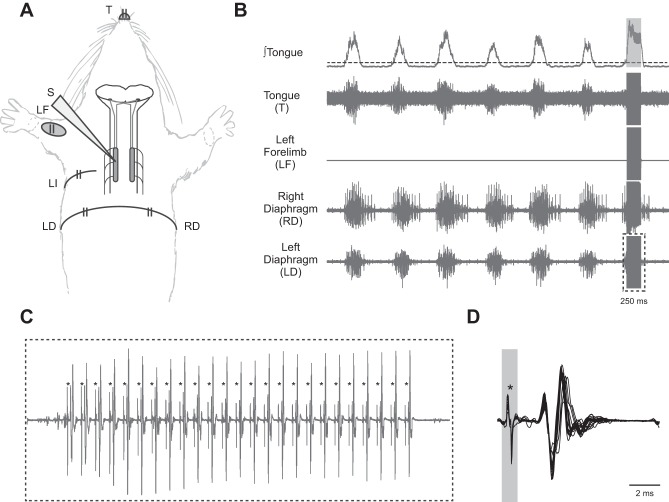
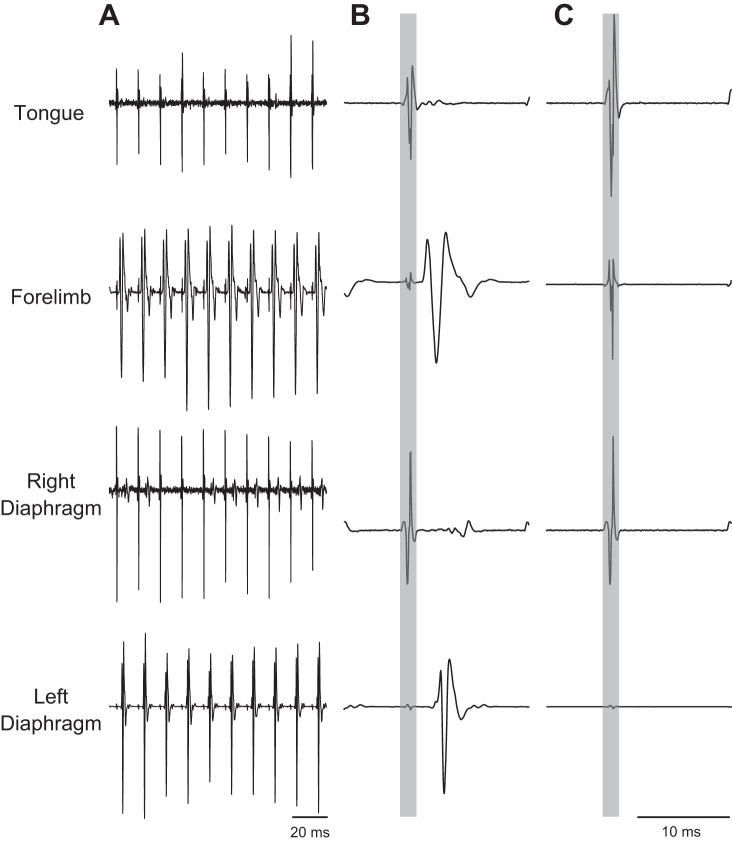
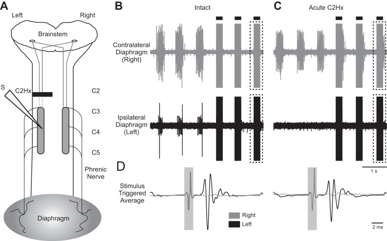
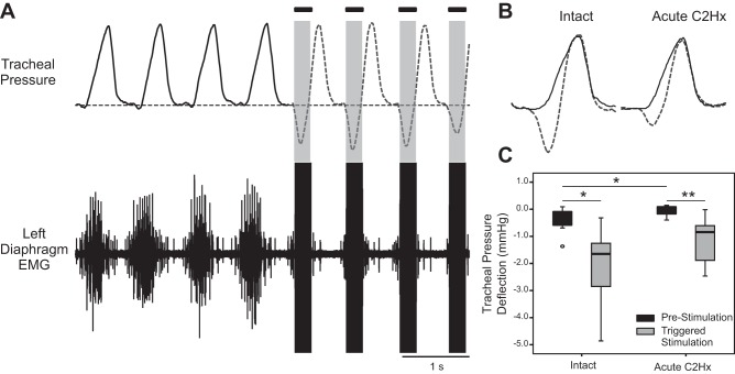
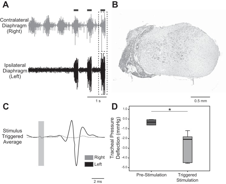
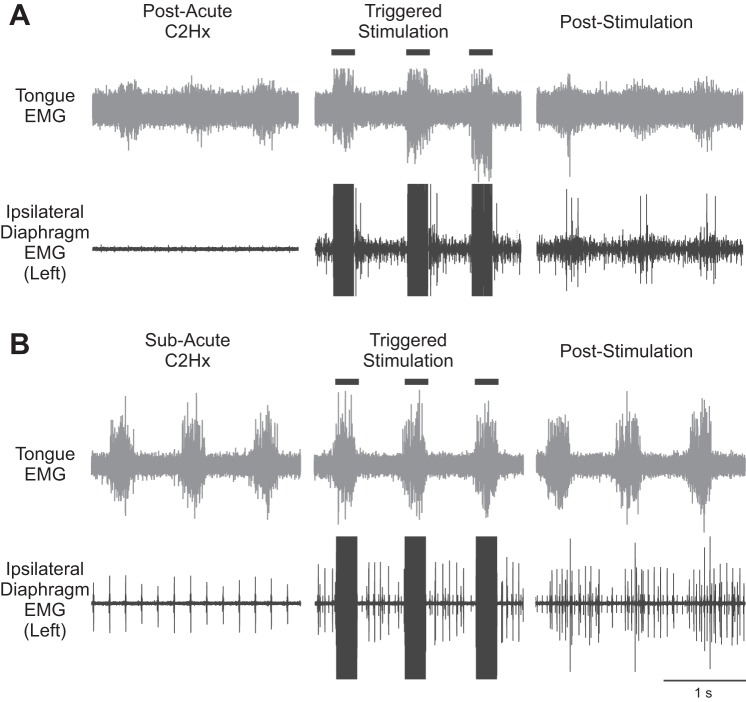
Intraspinal microstimulation (ISMS) using implanted electrodes can evoke locomotor movements after spinal cord injury (SCI) but has not been explored in the context of respiratory motor output. An advantage over epidural and direct muscle stimulation is the potential of ISMS to selectively stimulate components of the spinal respiratory network. The present study tested the hypothesis that medullary respiratory activity could be used to trigger midcervical ISMS and diaphragm motor unit activation in rats with cervical SCI. Studies were conducted after acute (hours) and subacute (5-21 days) C2 hemisection (C2Hx) injury in adult rats. Inspiratory bursting in the genioglossus (tongue) muscle was used to trigger a 250-ms train stimulus (100 Hz, 100-200 μA) to the ventral C4 spinal cord, targeting the phrenic motor nucleus. After both acute and subacute injury, genioglossus EMG activity effectively triggered ISMS and activated diaphragm motor units during the inspiratory phase. The ISMS paradigm also evoked short-term potentiation of spontaneous inspiratory activity in the previously paralyzed hemidiaphragm (i.e., bursting persisting beyond the stimulus period) in ∼70% of the C2Hx animals. We conclude that medullary inspiratory output can be used to trigger cervical ISMS and diaphragm activity after SCI. Further refinement of this method may enable "closed-loop-like" ISMS approaches to sustain ventilation after severe SCI.NEW & NOTEWORTHY We examined the feasibility of using intraspinal microstimulation (ISMS) of the cervical spinal cord to evoke diaphragm activity ipsilateral to acute and subacute hemisection of the upper cervical spinal cord of the rat. This proof-of-concept study demonstrated the efficacy of diaphragm activation, using an upper airway respiratory EMG signal to trigger ISMS at the level of the ipsilesional phrenic nucleus during acute and advanced postinjury intervals.
Keywords: diaphragm function; hypoglossal respiratory activity; phrenic motor nucleus; rat; respiration.
Copyright © 2017 the American Physiological Society.
Figures
Similar articles
-
Respiratory resetting elicited by single pulse spinal stimulation.Respir Physiol Neurobiol. 2020 Mar;274:103339. doi: 10.1016/j.resp.2019.103339. Epub 2019 Nov 14. Respir Physiol Neurobiol. 2020. PMID: 31734416 Free PMC article.
-
High-frequency epidural stimulation across the respiratory cycle evokes phrenic short-term potentiation after incomplete cervical spinal cord injury.J Neurophysiol. 2017 Oct 1;118(4):2344-2357. doi: 10.1152/jn.00913.2016. Epub 2017 Jun 14. J Neurophysiol. 2017. PMID: 28615341 Free PMC article.
-
Intraspinal microstimulation for respiratory muscle activation.Exp Neurol. 2018 Apr;302:93-103. doi: 10.1016/j.expneurol.2017.12.014. Epub 2018 Jan 2. Exp Neurol. 2018. PMID: 29305050 Free PMC article.
-
Adenosinergic mechanisms underlying recovery of diaphragm motor function following upper cervical spinal cord injury: potential therapeutic implications.Neurol Res. 2005 Mar;27(2):195-205. doi: 10.1179/016164105X21977. Neurol Res. 2005. PMID: 15829183 Review.
-
Intraspinal microstimulation for the recovery of function following spinal cord injury.Prog Brain Res. 2011;194:227-39. doi: 10.1016/B978-0-444-53815-4.00004-2. Prog Brain Res. 2011. PMID: 21867807 Free PMC article. Review.
Cited by
-
Spinal Interneurons as Gatekeepers to Neuroplasticity after Injury or Disease.J Neurosci. 2021 Feb 3;41(5):845-854. doi: 10.1523/JNEUROSCI.1654-20.2020. Epub 2021 Jan 20. J Neurosci. 2021. PMID: 33472820 Free PMC article. Review.
-
Respiratory resetting elicited by single pulse spinal stimulation.Respir Physiol Neurobiol. 2020 Mar;274:103339. doi: 10.1016/j.resp.2019.103339. Epub 2019 Nov 14. Respir Physiol Neurobiol. 2020. PMID: 31734416 Free PMC article.
-
Respiratory plasticity following spinal cord injury: perspectives from mouse to man.Neural Regen Res. 2022 Oct;17(10):2141-2148. doi: 10.4103/1673-5374.335839. Neural Regen Res. 2022. PMID: 35259820 Free PMC article. Review.
-
Contrasting Experimental Rodent Aftercare With Human Clinical Treatment for Cervical Spinal Cord Injury: Bridging the Translational "Valley of Death".J Neurotrauma. 2023 Dec;40(23-24):2469-2486. doi: 10.1089/neu.2023.0314. Epub 2023 Nov 10. J Neurotrauma. 2023. PMID: 37772694 Free PMC article. Review.
-
Role of Propriospinal Neurons in Control of Respiratory Muscles and Recovery of Breathing Following Injury.Front Syst Neurosci. 2020 Jan 17;13:84. doi: 10.3389/fnsys.2019.00084. eCollection 2019. Front Syst Neurosci. 2020. PMID: 32009911 Free PMC article. Review.
References
-
- Aertsen AM, Gerstein GL. Evaluation of neuronal connectivity: sensitivity of cross-correlation. Brain Res 340: 341–354, 1985. - PubMed
Publication types
MeSH terms
Grants and funding
LinkOut - more resources
Full Text Sources
Other Literature Sources
Medical
Miscellaneous