. 2016 Nov 23;36(47):11851-11864.
doi: 10.1523/JNEUROSCI.1700-16.2016.

Novel Roles for the Insulin-Regulated Glucose Transporter-4 in Hippocampally Dependent Memory

Affiliations

Novel Roles for the Insulin-Regulated Glucose Transporter-4 in Hippocampally Dependent Memory

Jiah Pearson-Leary et al. J Neurosci. .

Abstract

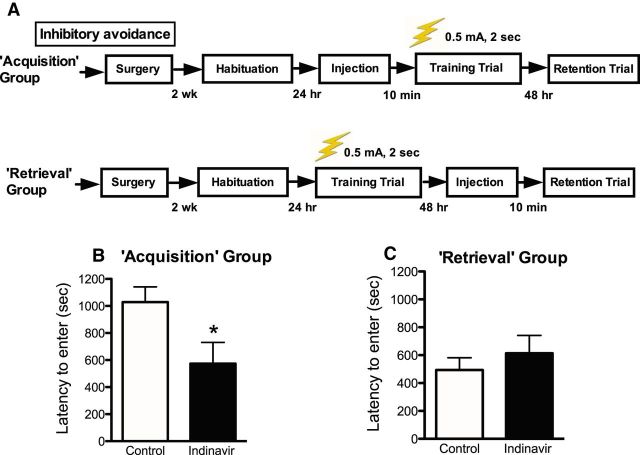
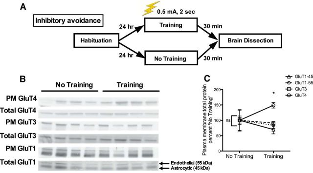
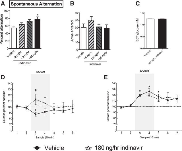
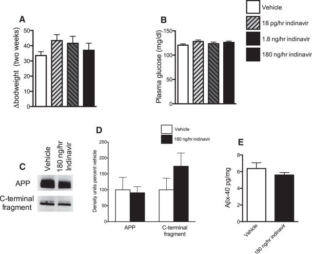
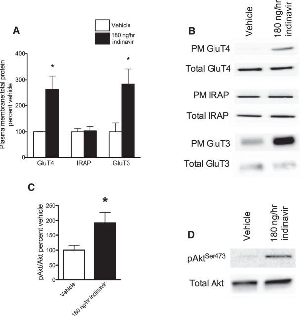
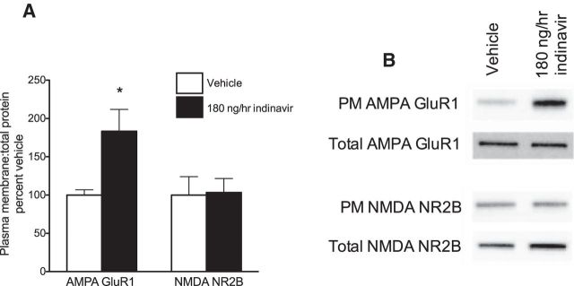
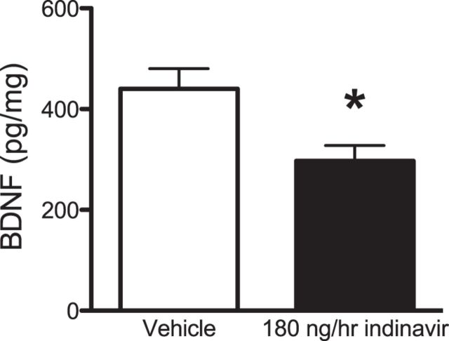
The insulin-regulated glucose transporter-4 (GluT4) is critical for insulin- and contractile-mediated glucose uptake in skeletal muscle. GluT4 is also expressed in some hippocampal neurons, but its functional role in the brain is unclear. Several established molecular modulators of memory processing regulate hippocampal GluT4 trafficking and hippocampal memory formation is limited by both glucose metabolism and insulin signaling. Therefore, we hypothesized that hippocampal GluT4 might be involved in memory processes. Here, we show that, in male rats, hippocampal GluT4 translocates to the plasma membrane after memory training and that acute, selective intrahippocampal inhibition of GluT4-mediated glucose transport impaired memory acquisition, but not memory retrieval. Other studies have shown that prolonged systemic GluT4 blockade causes insulin resistance. Unexpectedly, we found that prolonged hippocampal blockade of glucose transport through GluT4-upregulated markers of hippocampal insulin signaling prevented task-associated depletion of hippocampal glucose and enhanced both working and short-term memory while also impairing long-term memory. These effects were accompanied by increased expression of hippocampal AMPA GluR1 subunits and the neuronal GluT3, but decreased expression of hippocampal brain-derived neurotrophic factor, consistent with impaired ability to form long-term memories. Our findings are the first to show the cognitive impact of brain GluT4 modulation. They identify GluT4 as a key regulator of hippocampal memory processing and also suggest differential regulation of GluT4 in the hippocampus from that in peripheral tissues.

Significance statement: The role of insulin-regulated glucose transporter-4 (GluT4) in the brain is unclear. In the current study, we demonstrate that GluT4 is a critical component of hippocampal memory processes. Memory training increased hippocampal GluT4 translocation and memory acquisition was impaired by GluT4 blockade. Unexpectedly, whereas long-term inhibition of GluT4 impaired long-term memory, short-term memory was enhanced. These data further our understanding of the molecular mechanisms of memory and have particular significance for type 2 diabetes (in which GluT4 activity in the periphery is impaired) and Alzheimer's disease (which is linked to impaired brain insulin signaling and for which type 2 diabetes is a key risk factor). Both diseases cause marked impairment of hippocampal memory linked to hippocampal hypometabolism, suggesting the possibility that brain GluT4 dysregulation may be one cause of cognitive impairment in these disease states.

Keywords: GluT4; glucose; hippocampus; memory.


Figures

Figure 1.
Figure 1.
Inhibitory avoidance learning (0.5 mA shock, 2 s). A, Experimental outline for inhibitory avoidance procedures used to test the effects of intrahippocampal GluT4 inhibition on memory acquisition or retrieval (n = 8–10 per group). Graphs depict latencies for the retention trial. B, Intrahippocampal administration of the GluT4 inhibitor indinavir (200 ng dose) 10 min before the training trial (“Acquisition” group) decreased latencies to enter the dark chamber during the retention trial. C, Intrahippocampal administration of indinavir 10 min before day 2 testing (“Retrieval” group) had no effect on latencies to enter the dark chamber on retention trial testing (day 2 latencies are shown in the figure). Data are presented as the mean ± SEM; *p < 0.05.
Figure 2.
Figure 2.
Western blotting for glucose transporter expression in the hippocampus of rats killed immediately after the training trial of inhibitory avoidance. A, Experimental design outline. B, Western blot images from hippocampi of rats killed 30 min after the retention trial. For GluT1, the antibody used detected both endothelial (top band, 55 kDa) and astrocytic isoforms (bottom band, 45 kDa). C, There were no significant differences in PM glucose transporter expression between rats that were not tested for inhibitory avoidance (“No Training”). Rats that were killed 30 min after the training trial (“Training”) had increased PM GluT4 relative to PM GluT4 expression in untrained control rats. Inhibitory avoidance training did not lead to differences in expression of other hippocampal glucose transporters (n = 4–8 per group). Data are presented as the mean ± SEM; *p < 0.05.
Figure 3.
Figure 3.
Increased STM but impaired LTM after 2-week treatment with 180 ng/h indinavir. A, Experimental outline for object recognition testing. Separate cohorts of rats were tested 30 min (STM) or 16 h (LTM) after the 5 min training session (n = 8–13 per group). B, The 2 × 2 interaction was significant, with post hoc t tests showing increased discrimination index scores at 30 min (suggesting increased STM) and decreased scores at 16 h (suggesting decreased LTM). C, Experimental outline for contextual fear testing. Separate cohorts of rats were tested 30 min (STM) or 72 h (LTM) after the 3 min training session (n = 5–8 per group). D, Indinavir-treated rats had increased freezing behavior 30 min after the 3 min training session and decreased freezing behavior 72 h after the training session. E, There were no differences in freezing time in response to the tone cue. Data were analyzed statistically using a two-way ANOVA followed by Bonferroni's post hoc test and are presented as the mean ± SEM; *p < 0.05.
Figure 4.
Figure 4.
In vivo mD to measure markers of hippocampal metabolism while rats were engaged in a spatial working memory task. A, Two weeks of brain indinavir administration via intracerebroventricular Alzet minipump infusion led to a dose-dependent increase in spontaneous alternation behavior (n = 4–6 per group). B, Treatment did not affect total number of arms entered. C, Hippocampal glucose levels measured by in vivo mD showing no difference in hippocampal glucose levels after indinavir treatment. D, Indinavir prevented the task-associated dip in glucose that is well established to occur during spontaneous alternation testing (#p < 0.05 for comparison between vehicle and 180 ng/h indinavir at bin 3). E, Rats treated with vehicle or indinavir both showed significant increase in ECF lactate from baseline levels during SA testing (n = 9 per group for mD experiments). Data are presented as the mean ± SEM; *p < 0.05.
Figure 5.
Figure 5.
Measurements of peripheral metabolism and hippocampal AD-like pathology after central GluT4 inhibition. A, There was no change in bodyweight after 2 weeks of indinavir at any dose tested. B, There was no change in plasma glucose levels after 2 weeks of indinavir treatment (n = 5–7 per group). C, D, Representative Western blot images and graphs of total APP (C) and the intracellular C-terminal fragment of APP (D). There was no change in APP or C-terminal protein (n = 4–6 per group). E, ELISA data showing no change in Aβx-40 after indinavir treatment (n = 7–8 per group). Data are presented as the mean ± SEM; *p < 0.05.
Figure 6.
Figure 6.
GluT and PI3K activity in the hippocampus after 2 weeks of brain GluT4 inhibition (180 ng/h indinavir). A, Western blot demonstrating increased ratio of PM to total GluT4 in indinavir-treated rats. B, There was no effect of treatment on the GSV co-passenger IRAP. C, Ratio of phosphorylated Akt at serine 473 to total Akt was increased after indinavir treatment. D, Increase PM expression of GluT3 after indinavir treatment (n = 4–8 per group). Data are presented as the mean ± SEM; *p < 0.05.
Figure 7.
Figure 7.
Glutamate receptor expression in the hippocampus after 2 weeks of brain GluT4 inhibition. A, B, Ratio of PM to total AMPA GluR1 subunits were increased after indinavir treatment, but there was no change in the expression of NMDA NR2B subunits (n = 4–6 per group). Data are presented as the mean ± SEM; *p < 0.05.
Figure 8.
Figure 8.
BDNF expression in the hippocampus after 2 weeks of brain GluT4 inhibition. Indinivar treatment decreased the expression of hippocampal BDNF (n = 6 per group). Data are presented as the mean ± SEM; *p < 0.05.

References

    1. Alonso M, Vianna MR, Izquierdo I, Medina JH. Signaling mechanisms mediating BDNF modulation of memory formation in vivo in the hippocampus. Cell Mol Neurobiol. 2002a;22:663–674. doi: 10.1023/A:1021848706159. - DOI - PMC - PubMed
    1. Alonso M, Vianna MR, Depino AM, Mello e Souza T, Pereira P, Szapiro G, Viola H, Pitossi F, Izquierdo I, Medina JH. BDNF-triggered events in the rat hippocampus are required for both short- and long-term memory formation. Hippocampus. 2002b;12:551–560. doi: 10.1002/hipo.10035. - DOI - PubMed
    1. Alonso M, Bekinschtein P, Cammarota M, Vianna MR, Izquierdo I, Medina JH. Endogenous BDNF is required for long-term memory formation in the rat parietal cortex. Learn Mem. 2005;12:504–510. doi: 10.1101/lm.27305. - DOI - PMC - PubMed
    1. Alquier T, Leloup C, Lorsignol A, Pénicaud L. Translocable glucose transporters in the brain. Diabetes. 2006;55:S131–S138. doi: 10.2337/db06-S021. - DOI
    1. Anagnostaras SG, Gale GD, Fanselow MS. Hippocampus and contextual fear conditioning: recent controversies and advances. Hippocampus. 2001;11:8–17. - PubMed

Publication types

LinkOut - more resources