. 2016 Nov 23;8(366):366ra163.
doi: 10.1126/scitranslmed.aaf1090.

Two tissue-resident progenitor lineages drive distinct phenotypes of heterotopic ossification

Affiliations

Two tissue-resident progenitor lineages drive distinct phenotypes of heterotopic ossification

Devaveena Dey et al. Sci Transl Med. .

Abstract

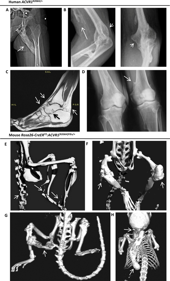
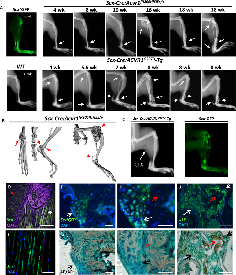
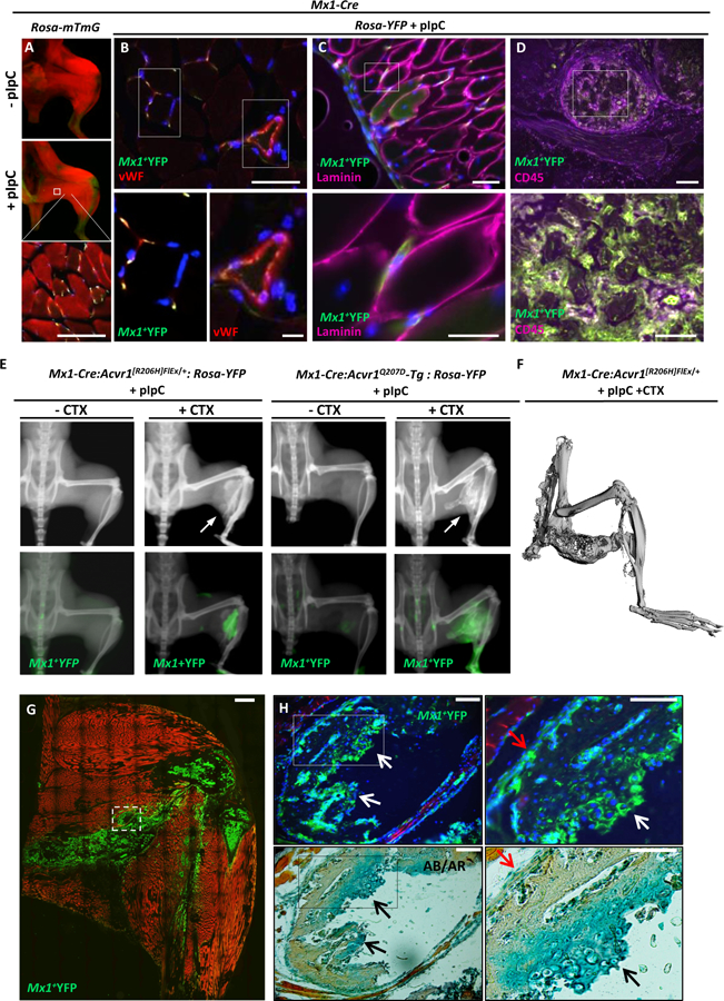
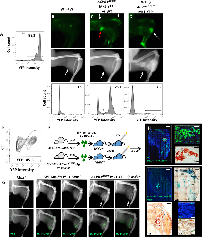
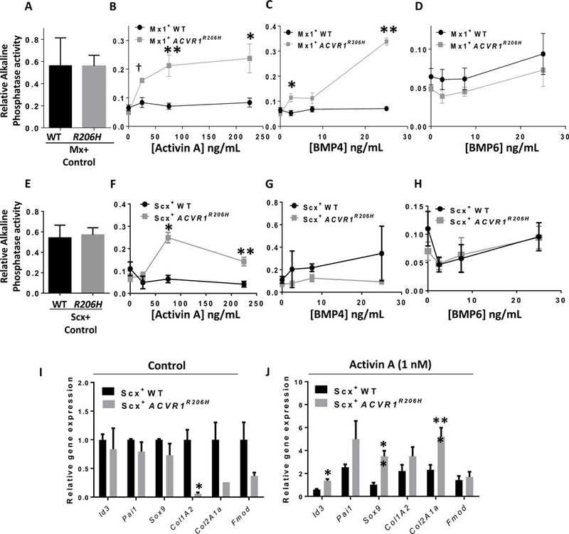
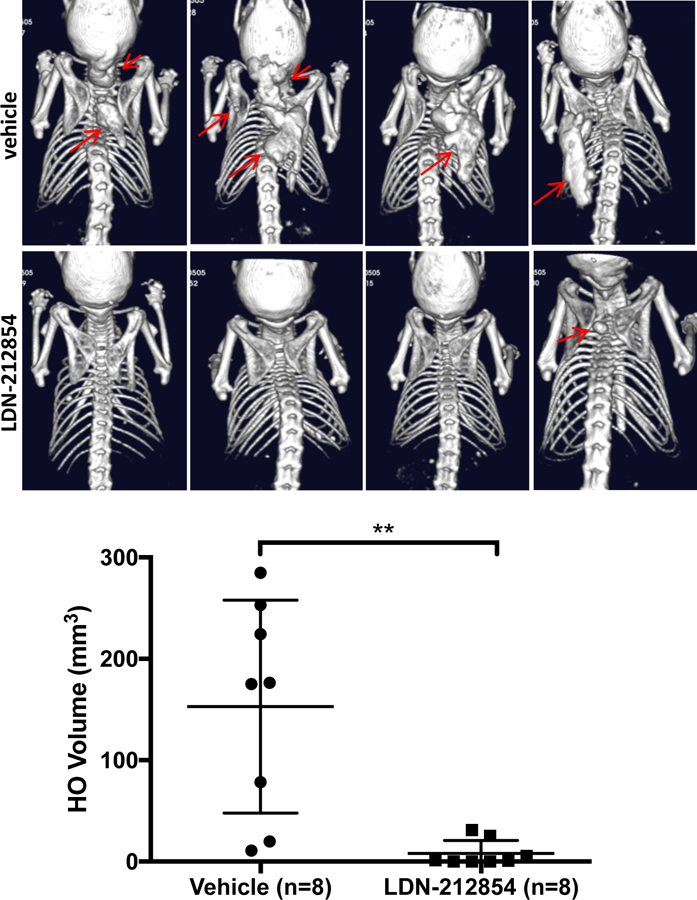
Fibrodysplasia ossificans progressiva (FOP), a congenital heterotopic ossification (HO) syndrome caused by gain-of-function mutations of bone morphogenetic protein (BMP) type I receptor ACVR1, manifests with progressive ossification of skeletal muscles, tendons, ligaments, and joints. In this disease, HO can occur in discrete flares, often triggered by injury or inflammation, or may progress incrementally without identified triggers. Mice harboring an Acvr1R206H knock-in allele recapitulate the phenotypic spectrum of FOP, including injury-responsive intramuscular HO and spontaneous articular, tendon, and ligament ossification. The cells that drive HO in these diverse tissues can be compartmentalized into two lineages: an Scx+ tendon-derived progenitor that mediates endochondral HO of ligaments and joints without exogenous injury, and a muscle-resident interstitial Mx1+ population that mediates intramuscular, injury-dependent endochondral HO. Expression of Acvr1R206H in either lineage confers aberrant gain of BMP signaling and chondrogenic differentiation in response to activin A and gives rise to mutation-expressing hypertrophic chondrocytes in HO lesions. Compared to Acvr1R206H, expression of the man-made, ligand-independent ACVR1Q207D mutation accelerates and increases the penetrance of all observed phenotypes, but does not abrogate the need for antecedent injury in muscle HO, demonstrating the need for an injury factor in addition to enhanced BMP signaling. Both injury-dependent intramuscular and spontaneous ligament HO in Acvr1R206H knock-in mice were effectively controlled by the selective ACVR1 inhibitor LDN-212854. Thus, diverse phenotypes of HO found in FOP are rooted in cell-autonomous effects of dysregulated ACVR1 signaling in nonoverlapping tissue-resident progenitor pools that may be addressed by systemic therapy or by modulating injury-mediated factors involved in their local recruitment.


Conflict of interest statement

Competing interests: ANE, SJH and LH are employees of Regeneron Pharmaceuticals, Inc. and hold stock in the company. PBY and Partners Healthcare hold patents on small molecules that inhibit BMP receptor signaling and may be entitled to royalties.

Figures

Fig 1.
Fig 1.. The classic FOP-causing ACVR1R206H allele is associated with intramuscular, peri-articular, ligament and tendon ossification in man and mouse.
(A-D) Radiographic manifestations in patients with ACVR1R206H/+ genotype-confirmed classic FOP. (A) Female patient with intramuscular ossification (arrow) bridging the ischium and left femoral shaft, infiltrating the gluteus and hamstring. (B) Male patient with intramuscular ossification infiltrating the biceps (long arrow) and dense peri-articular ossification surrounding the olecranon and coronoid fossae (short arrows). (C) T1 TSE MRI of a female patient reveals ossification of tibiotalar, talo-navicular and naviculo-calcaneal ligaments, and the Achilles tendon insertion (arrows) resulting in permanent plantar flexion. (D) A male patient with osteochondroma (arrow) of the distal humerus. (E-H) Micro-CT imaging of Rosa-CreERT2:Acvr1[R206H]FlEx/+ mice treated with tamoxifen for 10–12 wks reveals diffuse axial and appendicular heterotopic ossification after 4 wks, involving the paraspinal ligaments and Achilles tendons (E), peri-articular ossification of the knees (F), sporadic intramuscular ossification infiltrating hamstring and gastrocnemius, bridging ischium, femur and tibia (G), and of the interscapular muscles (H) with frequent handling for examination.
Fig 2.
Fig 2.. Expression of mutant ACVR1R206H or ACVR1Q207D alleles in Scx-lineage tendon-resident progenitor cells results in spontaneous peri-articular, ligament, and tendon ossification.
(A) Scleraxis expression is localized to tibiotalar ligament, patellar and Achilles tendons as observed by ex vivo fluorescence in Scx-GFP transgenic mice. Spontaneous ossification of tibiotalar ligament, patellar tendon and Achilles tendon progresses slowly in Scx-Cre:ACVR1R206H mice from 4 through 18 wks, with rapid progression in Scx-Cre:ACVR1Q207D-Tg mice from 4 through 8 weeks (B) Micro-CT of Scx-Cre:ACVR1R206H mice reveals distinct ossification of the tibiotalar ligament, Achilles tendon, and periarticular ossification, but no intramuscular ossification. (C-D) Scx-Cre:ACVR1Q207D-Tg:Rosa26-YFP mice injected with CTX (P21, gastrocnemius) develop typical tendon and ligament ossification, as seen by x-ray (P42) corresponding to areas of Scleraxis expression via Scx+YFP fluorescence but lack evidence of any intramuscular ossification (0/5 transgenic mice injected). (E) Cryosections of fibular head counterstained for CD45 (magenta) reveal Scleraxis expression (green) at ligamentous insertions (white arrow) but not in adjacent skeletal muscle (red arrow) in Scx-GFP transgenic mice. The HO lesions infiltrating the (F) talonavicular ligament, (G) talonavicular ligament in higher magnification, and (H) patellar tendon in Scx-Cre:ACVR1Q207D-Tg;Rosa26-YFP mice reflect the contribution of Scx+YFP+ cells to nearly all Alcian Blue (AB)-stained hypertrophic chondrocytes and heterotopic cartilage (white arrows) in these lesions, but essentially no contribution to osteocytes or mineralized matrix (red arrows) stained with Alizarin Red (AR), with DAPI as a nuclear counter-stain.
Fig 3.
Fig 3.. Expression of mutant ACVR1R206H or ACVR1Q207D alleles in Mx1-lineages results in injury-dependent intramuscular heterotopic ossification
(A) Treatment of Mx1-Cre:Rosa26-mTmG (mTmG) mice with pIpC activates GFP expression within skeletal muscle interstitial cells (red=mTomato, yellow=mGFP). (B-D) Gastrocnemius muscles of pIpC treated Mx1-Cre:Rosa26-YFP mice reveals a portion of Mx1+ lineage (YFP, green) cells in the interstitium and microvascular endothelium co-staining with DAPI (blue) and vWF (red) and shown at higher magnification (B, and inset panels below). Mx1+ cells were located consistently outside of myofiber basement membranes (C), based on laminin staining (magenta), and accounted for a high percentage of bone marrow cells based on co-staining with CD45 (magenta, D). (E) Treatment of Mx1-Cre:Acvr1[R206H]FlEx/+:Rosa26-YFP and Mx1-Cre:ACVR1Q207D-Tg:Rosa26-YFP mice with pIpC results in injury-dependent ossification of hindlimb muscles following cardiotoxin (CTX) mediated injury, with YFP-marked intramuscular bone. (F) Micro-CT imaging of Mx1-Cre:Acvr1[R206H]FlEx/+ mice treated with pIpC and CTX reveals ossification infiltrating hamstring and gastrocnemius, fusing ischium and tibia without articular involvement. (G) Intramuscular HO in Mx1-Cre:ACVR1Q207D-Tg:Rosa26-mTmG mice is derived from GFP-marked Mx1+ cells, which also marks heterotopic bone marrow, with detail of lesion structure shown (G) and at higher magnification (H) revealing a mutant Mx1+ origin for nearly 100% of Alcian Blue (AB)-stained hypertrophic chondrocytes, and periosteal cells, in contrast to no contribution to Alizarin Red (AR)-stained mineralized matrix or associated osteocytes.
Fig 4.
Fig 4.. Mx1-lineage muscle interstitial but not bone marrow cells are sufficient for intramuscular HO.
(A-D) Reciprocal bone marrow transplant experiments demonstrate medullary expression of mutant ACVR1 is dispensable for HO. (A) Control Mx1-Cre:Rosa26-YFP or mutant Mx1-Cre:ACVR1Q207D-Tg:Rosa26-YFP mice treated with pIpC exhibited >90% YFP labeling of marrow cells. Control or mutant mice were preconditioned antenatally with busulfan (E16, maternal i.p. injection), and engrafted at P2 with total bone marrow cells (5 ×10⁵ cells i.p.) harvested at P21 from WT or mutant donor mice previously treated with pIpC (P7-P21). In contrast to WT mice engrafted with WT marrow (B), WT mice engrafted with mutant marrow (C) exhibited a high percentage of YFP+ bone marrow comparable to mutant donor mice (A) by flow cytometry 78.2% ± 15.9% (n=5), and ex vivo fluorescence (white arrows), but did not exhibit injury-dependent ossification following CTX, despite the presence of infiltrating YFP+ cells due to CTX-induced inflammation (red arrow). Conversely, Mx1-Cre:ACVR1Q207D-Tg:Rosa26-YFP mice engrafted with WT marrow (D) exhibited very low frequencies of residual YFP+ bone marrow by flow cytometry (bottom panel, 0.5–5%; n=11), but exhibited robust CTX-induced intramuscular ossification with YFP+ lesions (white arrows, top and middle panel). (E-I) Mx1+ lineage muscle interstitial cell engraftment studies demonstrate Mx1+ lineage interstitial cells are sufficient for injury-dependent intramuscular HO. Mx1+YFP+ (5×10⁵) cells sorted from the muscles of P21 control Mx1-Cre:Rosa26-YFP or mutant Mx1-Cre:ACVR1Q207D-Tg:Rosa26-YFP mice previously treated with pIpC (P7-P19) were transplanted into gastrocnemius muscles of Dmdmdx−5cv:Rag1null (Mdx−/−) mice (P21) in Matrigel (E-F). In comparison to Mdx−/− control mice injected with Matrigel only (G, left), Mdx−/− mice injected with WT Mx1+YFP+ cells exhibited engraftment after 6 wks based on YFP fluorescence but no heterotopic ossification with or without injury (G, middle panel, no lesions seen in 5 treated mice), whereas Mdx−/− mice injected with mutant Mx1+YFP+ cells exhibited engraftment and developed intramuscular ossification following CTX treatment (G, right panel, lesions seen in 3/5 mice treated). (H) Histological analysis of mice injected with WT Mx1+YFP+ control cells demonstrated engraftment of YFP+ cells interspersed in gastrocnemius and popliteal fossa, all of which stain with Oil Red O (ORO). (I) Mice engrafted with ACVR1Q207D Mx1+YFP+ cells demonstrated engraftment of YFP+ cells throughout HO lesions of the gastrocnemius, with mineralization evident by Alizarin Red (AR), formation of ectopic cartilage demonstrated by Alcian Blue (AB), but were notable for the absence of YFP fluorescence in heterotopic marrow, shown by fluorescence and DAPI counter-staining (inset top panel), and the co-localization of YFP fluorescence with mineralized areas (AR, inset bottom panel).
Fig 5.
Fig 5.. Expression of ACVR1R206H in Mx1 and Scx-lineage cells modifies their osteo-chondrogenic differentiation potential in a ligand-dependent manner.
(A) Spontaneous (n=8) and (B-D) ligand-induced alkaline phosphatase expression in freshly isolated interstitial Mx1+ACVR1R206H and Mx1+ control cells obtained from quiescent, non-injured muscles following culture in the presence or absence of ligand for 4 d (n=4; p-values from left to right: 5B. †0.006,**0.01, *0.03; 5C. *0.02, **2×10⁻⁶). (E) Spontaneous (n=4) and (F-H) ligand-induced alkaline phosphatase expression in freshly isolated YFP+ cells from 2-week old Scx-Cre:ACVR1R206H-Tg:Rosa-YFP (Scx+ACVR1R206H) and Scx-Cre:Rosa26-YFP (Scx+WT) mice following culture in the presence or absence of ligand for 4 d (n=4; p-values from left to right: *1.3×10⁻⁶, **0.0001). Expression of BMP and TGF-β transcriptional targets and endochondral genes in Scx+ACVR1R206H and Scx+WT cells without (I) and with (J) Activin A (1 nM) treatment for 48 h; (n=4; *p<0.02, **p<0.02, ***p<0.003). Unpaired Student’s t-test was used for statistical analysis.
Fig 6.
Fig 6.. Treatment with an ACVR1-selective kinase inhibitor prevents HO in global knock-in Acvr1R206H mice in vivo.
Rosa-CreERT2:Acvr1[R206H]FlEx/+mice treated with tamoxifen for 10–12 wks, followed by treatment with vehicle for 4 wks developed spontaneous joint and ligamentous HO, as well as prominent interscapular HO at sites of handling and examination (red arrows, top panels), whereas animals treated with the ACVR1-selective inhibitor LDN-212854 (2.5 mg/kg twice daily s.c.) demonstrated near-complete inhibition of joint, ligamentous, and interscapular HO (red arrow, lower panel).

Similar articles

Cited by

References

    1. Kaplan FS, Le Merrer M, Glaser DL, Pignolo RJ, Goldsby RE, Kitterman JA, Groppe J, Shore EM, Fibrodysplasia ossificans progressiva. Best Pract Res Clin Rheumatol 22, 191–205 (2008). - PMC - PubMed
    1. Pignolo RJ, Bedford-Gay C, Liljesthrom M, Durbin-Johnson BP, Shore EM, Rocke DM, Kaplan FS, The Natural History of Flare-Ups in Fibrodysplasia Ossificans Progressiva (FOP): A Comprehensive Global Assessment. J Bone Miner Res 31, 650–656 (2016). - PMC - PubMed
    1. Shore EM, Xu M, Feldman GJ, Fenstermacher DA, Brown MA, Kaplan FS, A recurrent mutation in the BMP type I receptor ACVR1 causes inherited and sporadic fibrodysplasia ossificans progressiva. Nat Genet 38, 525–527 (2006). - PubMed
    1. Kaplan FS, Xu M, Seemann P, Connor JM, Glaser DL, Carroll L, Delai P, Fastnacht-Urban E, Forman SJ, Gillessen-Kaesbach G, Hoover-Fong J, Koster B, Pauli RM, Reardon W, Zaidi SA, Zasloff M, Morhart R, Mundlos S, Groppe J, Shore EM, Classic and atypical fibrodysplasia ossificans progressiva (FOP) phenotypes are caused by mutations in the bone morphogenetic protein (BMP) type I receptor ACVR1. Human mutation 30, 379–390 (2009). - PMC - PubMed
    1. Fukuda T, Kohda M, Kanomata K, Nojima J, Nakamura A, Kamizono J, Noguchi Y, Iwakiri K, Kondo T, Kurose J, Endo K, Awakura T, Fukushi J, Nakashima Y, Chiyonobu T, Kawara A, Nishida Y, Wada I, Akita M, Komori T, Nakayama K, Nanba A, Maruki Y, Yoda T, Tomoda H, Yu PB, Shore EM, Kaplan FS, Miyazono K, Matsuoka M, Ikebuchi K, Ohtake A, Oda H, Jimi E, Owan I, Okazaki Y, Katagiri T, Constitutively activated ALK2 and increased SMAD1/5 cooperatively induce bone morphogenetic protein signaling in fibrodysplasia ossificans progressiva. J Biol Chem 284, 7149–7156 (2009). - PMC - PubMed

MeSH terms