. 2016 Nov 24:6:37516.
doi: 10.1038/srep37516.

Wide-Field Detected Fourier Transform CARS Microscopy

Affiliations

Wide-Field Detected Fourier Transform CARS Microscopy

Alex Soares Duarte et al. Sci Rep. .

Abstract

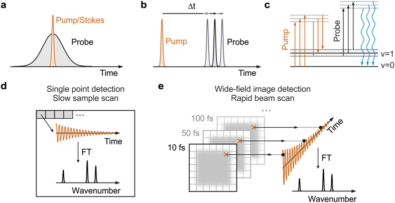
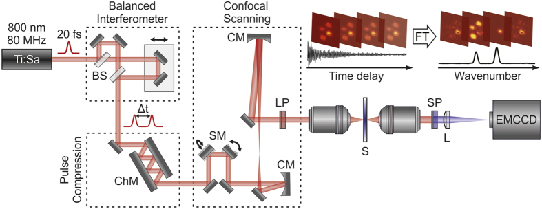
We present a wide-field imaging implementation of Fourier transform coherent anti-Stokes Raman scattering (wide-field detected FT-CARS) microscopy capable of acquiring high-contrast label-free but chemically specific images over the full vibrational 'fingerprint' region, suitable for a large field of view. Rapid resonant mechanical scanning of the illumination beam coupled with highly sensitive, camera-based detection of the CARS signal allows for fast and direct hyperspectral wide-field image acquisition, while minimizing sample damage. Intrinsic to FT-CARS microscopy, the ability to control the range of time-delays between pump and probe pulses allows for fine tuning of spectral resolution, bandwidth and imaging speed while maintaining full duty cycle. We outline the basic principles of wide-field detected FT-CARS microscopy and demonstrate how it can be used as a sensitive optical probe for chemically specific Raman imaging.


Figures

Figure 1
Figure 1. Overview of coherent anti-Stokes Raman scattering (CARS) microscopy.
(a) Pulse-timing diagram for broadband frequency-domain CARS and (b) time-domain CARS. (c) Wave-mixing energy ladder diagram describing the generation of blue-shifted CARS photons after three electric field interactions with the sample. Fourier transform CARS microscopy relying on single point (d) and wide-field image (e) detection. CARS spectra are retrieved after Fourier transformation (FT) of the obtained coherent oscillations.
Figure 2
Figure 2. Wide-field detected FT-CARS microscopy setup scheme and concept.
Combination of a balanced interferometer, chirped mirror compression and reflective beam scanning delivers 18 fs pulses to the focus of a high NA microscope objective. Complete images are read out for each interpulse time delay before being Fourier transformed to produce a hyperspectral image. BS – beamsplitter, ChM – chirped mirrors, SM – scanning mirrors, LP – long pass filter, SP – short pass filter, S - sample, L - tube lens, CM - curved mirror.
Figure 3
Figure 3. Wide-field detected FT-CARS microscopy on toluene.
(a) Time-independent coherent anti-Stokes background scattering of the scanning region (30 × 30 μm2). The inhomogeneous intensity distribution arises from the harmonic motion of the scanning mirrors. (b) Coherent oscillations of toluene recorded for 3.5 ps on a single pixel (dashed circle in a). The data was recorded in 30 s with an EMCCD camera integration time of 20 ms in the absence of EM gain. (c) Fourier power spectrum of the coherent oscillations in (a) showing the characteristic vibrational peaks of toluene. (d) Fourier power map along the vertical dashed black line in (a). We note, that the Fourier intensity scales quadratically with the incident power (inset).
Figure 4
Figure 4. Wide-field detected FT-CARS microscopy applied to a dry mixture of PMMA and PS beads.
Normalized Fourier power map (9 × 9 μm2) at (a) 810 cm−1 and (b) 1010 cm−1 corresponding to the dominant low-frequency modes identifying PMMA and PS beads, respectively. (c) Fourier power map at 1270 cm−1 characterizing the noise level of the measurement (normalized by the 810 cm−1 peak). (d) Composite images of (a,b) illustrating the chemical sensitivity of wide-field detected FT-CARS microscopy. (e) Top: Normalized Fourier power spectrum of PMMA bead pointed by the arrow in (a) Bottom: normalized Fourier power spectrum of a the PS bead pointed by the arrow in (b).

Similar articles

Cited by

References

    1. Camp C. H. Jr & Cicerone M. T. Chemically sensitive bioimaging with coherent Raman scattering. Nat. Photonics 9, 295–305 (2015).
    1. Cheng J.-X. & Xie X. S. Vibrational spectroscopic imaging of living systems: An emerging platform for biology and medicine. Science (80-.). 350, aaa8870 (2015). - PubMed
    1. Evans C. L. & Xie X. S. Coherent Anti-Stokes Raman Scattering Microscopy: Chemical Imaging for Biology and Medicine. Annu. Rev. Anal. Chem. 1, 883–909 (2008). - PubMed
    1. Krafft C., Dietzek B. & Popp J. Raman and CARS microspectroscopy of cells and tissues. Analyst 134, 1046–1057 (2009). - PubMed
    1. Rez P. et al.. Damage-free vibrational spectroscopy of biological materials in the electron microscope. Nat. Commun. 7, 10945 (2016). - PMC - PubMed

Publication types

LinkOut - more resources