. 2016 Nov 10:22:1318-1331.
eCollection 2016.

Genetic and immunohistochemical analysis of HSPA5 in mouse and human retinas

Affiliations

Genetic and immunohistochemical analysis of HSPA5 in mouse and human retinas

Sumana R Chintalapudi et al. Mol Vis. .

Abstract

Purpose: Photoreceptor degenerative diseases are among the leading causes of vision loss. Although the causative genetic mutations are often known, mechanisms leading to photoreceptor degeneration remain poorly defined. We have previously demonstrated that the photoreceptor membrane-associated protein XAP-1 antigen is a product of the HSPA5 gene. In this study, we used systems genetic methods, statistical modeling, and immunostaining to identify and analyze candidate genes that modulate Hspa5 expression in the retina.

Methods: Quantitative trait locus (QTL) mapping was used to map the genomic region that regulates Hspa5 in the cross between C57BL/6J X DBA/2J mice (BXD) genetic reference panel. The stepwise refinement of candidate genes was based on expression QTL mapping, gene expression correlation analyses (direct and partial), and analysis of regional sequence variants. The subcellular localization of candidate proteins and HSPA5 in mouse and human retinas was evaluated by immunohistochemistry. Differences in the localization of extracellular HSPA5 were assessed between healthy human donor and atrophic age-related macular degeneration (AMD) donor eyes.

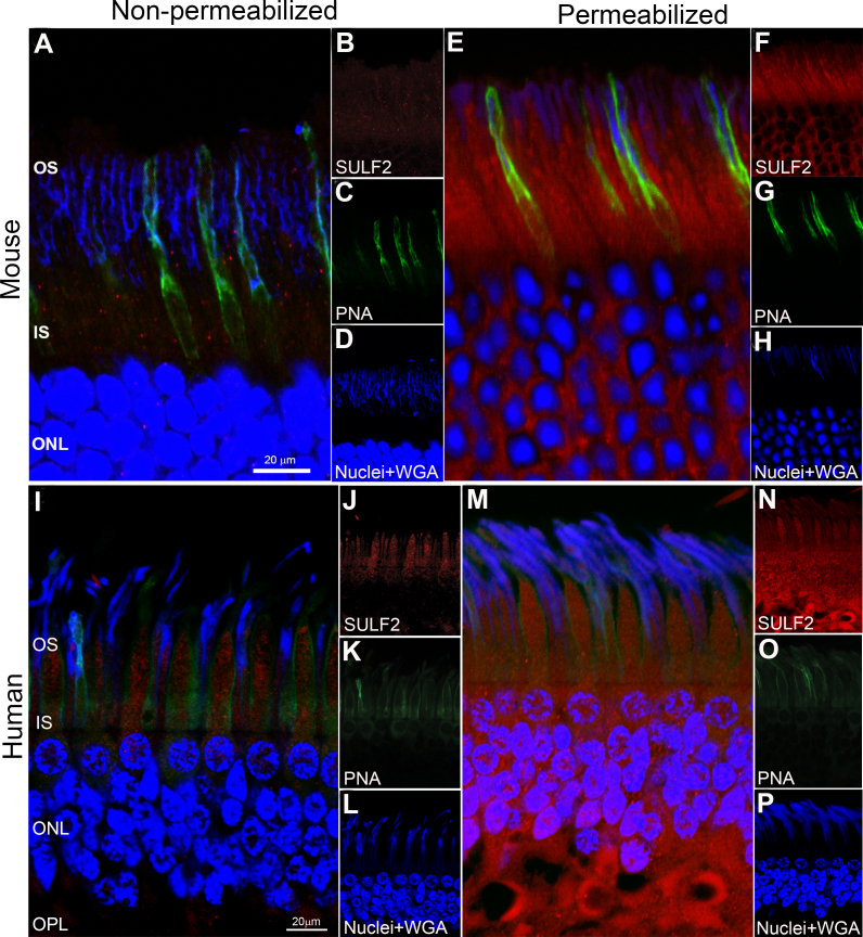
Results: In the eyes of healthy mice, extracellular HSPA5 was confined to the area around the cone photoreceptor outer segments. Mapping variation in Hspa5 mRNA expression levels in the retina revealed a statistically significant trans-acting expression QTL (eQTL) on Chromosome 2 (Chr 2) and a suggestive locus on Chr 15. Sulf2 on Chr 2 was the strongest candidate gene based on partial correlation analysis, Pearson correlation with Hspa5, expression levels in the retina, a missense variant in exon 14, and its reported function in the extracellular matrix and interphotoreceptor matrix. SULF2 is localized to the rod and cone photoreceptors in both human and mouse retinas. In human retinas with no pathology, extracellular HSPA5 was localized around many cones within the macular area. In contrast, fewer HSPA5-immunopositive cones were observed in the retinas from AMD donors.

Conclusions: We identified Sulf2 as a candidate gene modulating the Hspa5 expression in the retina. The preferential loss of HSPA5 in the interphotoreceptor matrix around cone photoreceptors in atrophic AMD retinas opens up new avenues for exploring the changes in interphotoreceptor matrix (IPM) that are associated with macular disease.


Figures

Figure 1
Figure 1
Immunohistochemical localization of HSPA5 in mice retinas (en face presentation). A-D: HSPA5 (green) co-localizes with PNA (red) in the IPM surrounding the cone photoreceptors. The co-localization of HSPA5 and PNA is shown in yellow (white arrow). A minimal amount of HSPA5 immunolabeling is associated with WGA around rods (black arrow). Binding of WGA to the IPM surrounding the rods is shown in blue. Scale bar = 10 µm.
Figure 2
Figure 2
Localization of intracellular and extracellular HSPA5 in retina cross sections from mice. A-D: In non-permeabilized sections, HSPA5 (green) co-localizes with PNA (red) in the IPM surrounding the cone photoreceptors. The co-localization of HSPA5 and PNA is shown in yellow. E-H: In permeabilized retina cross sections, HSPA5 co-localizes with PNA in a pattern similar to the non-permeabilized retina cross sections (yellow). OS = outer segments, IN = inner segments, ONL = outer nuclear layer. Scale bar = 20 µm.
Figure 3
Figure 3
Expression levels of Hspa5 across the BXD strains and simple interval mapping. A: Rank-ordered mean Hspa5 levels across the BXD recombinant inbred family ranges from 6.9±0.2 in BXD80 to 8.4±0.3 in BXD102. The values denote the normalized relative expression levels on a log2 scale (mean±SEM). B: The QTL simple interval map of genomic regions that modulate Hspa5 expression in the retinas of BXD mice. A significant trans-eQTL for Hspa5 is present on Chr 2 and a second trans-eQTL is present on Chr 15 (circles). The blue tracings indicate the LRS scores across the genome. The x-axis represents the LRS scores across the genome. The horizontal lines represent the transcript-specific significance thresholds for significant (LRS = 17.21) and suggestive (LRS = 10.48) LRS levels indicated by red and gray horizontal lines, respectively. The filled triangle on Chr 2 indicates the location of Hspa5 in the genome.
Figure 4
Figure 4
Heat map analysis of Hspa5 covariates and candidate genes. A: Heat map of the top 100 covariates of Hspa5 as determined by the Pearson correlation analysis. Probe sets for the various genes represent columns and the genomic location for each chromosome represents rows. Tight bands of correlates are present on Chr 2 and Chr 15 at the location of the QTL peaks, which were revealed by simple interval mapping. B: Heat map containing B4galt5, Dpm1, Fbln1, Glt8d3, Sulf2, Pmm1, and Hspa5. The hue intensity indicates the strength of the association between gene expression and genomic location. The red and blue colors indicate that the D or B allele increases trait values, respectively. The orange triangles show the location of each gene on the chromosomes. B4galt5, Dpm1, Fbln1, Glt8d3, Sulf2, and Pmm1 show a strong consistency of genetic regulation across mRNA regions on Chr 2 and Chr 15 (arrows).
Figure 5
Figure 5
Immunohistochemical detection of SULF2 in non-pathological mouse and human donor retina cross sections. A-D: In non-permeabilized mouse retina cross sections, SULF2 (red) is observed in the extracellular space around the photoreceptor inner and outer segments. E-H: In permeabilized mouse retina cross sections, SULF2 (red) is predominantly observed intracellularly in the rod (blue) and cone (green) photoreceptors, as well as in the cell bodies. I-L: In non-permeabilized non-pathological human retina cross sections, the labeling pattern of SULF2 (red) is identical to that observed in mice. M-P: In permeabilized human retina cross sections, the SULF2 (red) labeling pattern is also identical to that observed in mice. OS = outer segments, IN = inner segments, ONL = outer nuclear layer, OPL = outer plexiform layer. Scale bar = 20 µm.
Figure 6
Figure 6
HSPA5 co-localizes with PNA in flat mounts and cross sections of the human macula with no pathology. A-D: In retinal flat mounts, HSPA5 (green) co-localizes with PNA (red). Punctate yellow indicates co-localization of HSPA5 and PNA in the IPM of the cone photoreceptors (white arrow). WGA (blue) indicates the IPM of the rods (black arrow). E-H: In retina cross sections, HSPA5 (green) also co-localizes with PNA along the full length of the cones (red, white arrows). WGA (blue) indicates the IPM of the rods. Scale bar = 10 µm. Electron microscopic immunohistochemical localization of HSPA5 (gold particles) in human retinas (with no ocular pathology). I: HSPA5 is localized to the flocculent material in the IPM surrounding the cone outer segments between adjacent cone and rod outer segments (box). Scale bar = 2 µm. J: Higher magnification view of HSPA5 localization (black arrow; the boxed area in G is shown magnified in H). COS = cone outer segment, CIS = cone inner segment, and ROS = rod outer segment. Scale bar = 200 nm. Images are representative of all the donor eyes that were used in the study.
Figure 7
Figure 7
Localization of HSPA5 and WGA in retinas from human donors with no ocular pathology and age-matched donors with diagnosis of atrophic AMD. A-F: Localization of HSPA5 and WGA in the retina of a 77-year-old male who had cataract surgery with intraocular lens (IOL) insertion and no history of AMD. A-C: In the central macular region of a healthy donor retina, the merged image shows the localization of HSPA5 (green) to the cone IPM in the fovea with lesser labeling around the rod photoreceptors (blue). D-F: Merged image of the peripheral retinal area from the same healthy donor demonstrates scattered labeling of HSPA5 (green) on the cone and rod photoreceptors (blue). G-L: Localization of HSPA5 and WGA in the retina of a 91-year-old female with dry AMD and bilateral cataracts. G-I: Merged image of the central macular region from a dry AMD donor retina shows negligible HSPA5 (green) staining. J-L: Merged image of the peripheral retinal area from the same donor with dry AMD shows scattered and unstructured HSPA5 (green) staining. Scale bar = 10 µm. Images are representative of all the donor eyes that were used in the study.

Similar articles

Cited by

References

    1. Beere HM, Wolf BB, Cain K, Mosser DD, Mahboubi A, Kuwana T, Tailor P, Morimoto RI, Cohen GM, Green DR. Heat-shock protein 70 inhibits apoptosis by preventing recruitment of procaspase-9 to the Apaf-1 apoptosome. Nat Cell Biol. 2000;2:469–75. - PubMed
    1. Delpino A, Castelli M. The 78 kDa glucose-regulated protein (GRP78/BIP) is expressed on the cell membrane, is released into cell culture medium and is also present in human peripheral circulation. Biosci Rep. 2002;22:407–20. - PubMed
    1. Ni M, Zhang Y, Lee AS. Beyond the endoplasmic reticulum: atypical GRP78 in cell viability, signalling and therapeutic targeting. Biochem J. 2011;434:181–8. - PMC - PubMed
    1. Ni M, Lee AS. ER chaperones in mammalian development and human diseases. FEBS Lett. 2007;581:3641–51. - PMC - PubMed
    1. Sun FC, Wei S, Li CW, Chang YS, Chao CC, Lai YK. Localization of GRP78 to mitochondria under the unfolded protein response. Biochem J. 2006;396:31–9. - PMC - PubMed

LinkOut - more resources