The influence of exendin-4 intervention on -obese diabetic mouse blood and the pancreatic tissue immune microenvironment
- PMID: 27882092
- PMCID: PMC5103724
- DOI: 10.3892/etm.2016.3694
The influence of exendin-4 intervention on -obese diabetic mouse blood and the pancreatic tissue immune microenvironment
Abstract
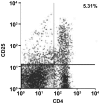
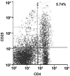
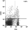
The aim of the study was to determine the influence of exendin-4 intervention on non-obese diabetic (NOD) mouse blood and the pancreatic tissue immune microenvironment. A total of 40 clean NOD mice were used in the study and randomly divided into 4 groups (n=10/group). The first group was blank control group D with normal saline intervention, and with different doses of exendin, i.e.,-4 2, 4 and 8 µg/kg/day. The three remaining groups were: i) Low-dose group A; ii) medium-dose group B; and iii) high-dose group C. Mice in the four groups went through intervention for 8 weeks. Their mass and blood glucose levels were tested each week. After 8 weeks, the mice were sacrificed, and mouse serum samples were reserved. The ELISA method was used to test peripheral blood (PB), IL-2, IFN-γ and IL-10 levels. Pancreatic samples were created. Immunohistochemistry was used to observe the infiltration degree of mouse pancreatitis and the local expression state of pancreatic IL-10. Mouse pancreatic tissues were suspended in pancreatic cell suspension. Flow cytometry was used to test the state of T-cell subsets CD4 and CD25. Mouse pancreatitis in control group D was mainly at grade 2and 3. Under a light microscope, it was observed that pancreatic cell morphology was in disorder, and the size and quantity of the pancreas was small. Mouse pancreatitis in the exendin-4 low-dose group A, medium-dose group B and high-dose group C was mainly at grade 0 and 1. Under a light microscope, it was observed that pancreatic cell morphology improved, the infiltration degree of lymphocyte was improved and pancreatic islet size was restored somewhat. Additionally, a few brownish granules were identified within the pancreatic sample cells in control group D. There were many brownish granules with deep color within the pancreatic sample cells in exendin-4 low-dose group A, medium-dose group B and high-dose group C. IL-10 immunohistochemistry scores in the low-dose group A, medium-dose group B and high-dose group C were 3.82±0.72, 4.34±0.86 and 4.81±0.94, respectively, and were higher than the score of 2.25±0.63 in control group D. CD4+CD25+T-cell proportions in mouse pancreatic tissues of low-dose group A, medium-dose group B and high-dose group C were 5.31, 5.53 and 5.74%, respectively, which were higher than that of the CD4+CD25+T-cell proportion (1.62% in control group D). The CD4+CD25high T-cell proportion in CD4+T-cells in group A, B and C increased. Compared with control group D, serum IL-10 levels in the exendin-4 low-dose group A, medium-dose group B and high-dose group C increased (P<0.05), while levels of IL-2 and IFN-γ decreased (P<0.05). Additionally, the difference of serum IL-10, IL-2 and IFN-γ levels in the low-dose group A, medium-dose group B and high-dose group C was of statistical significance (P<0.05). Exendin-4 intervention can increase quantities of CD4 and CD8+T cells in NOD mouse pancreases, with PB IL-10 expression and local expression of IL-10 in pancreatic tissues. It also can inhibit the expression of serum IL-2 and IFN-γ, regulate the organism immune microenvironment and prevent diabetes. CD4+CD25high T cells increase in NOD tumor infiltration lymphocytes mediated by exendin-4 intervention, which may be related to the fact that exendin-4 inhibits the lethal effect of CD8+T cells through contact among cells and eventually exerts immunosuppressive effect.
Keywords: NOD mice; diabetes; exendin-4; immune microenvironment.
Figures


References
-
- Lind K, Hühn MH, Flodström-Tullberg M. Immunology in the clinic review series; focus on type 1 diabetes and viruses: The innate immune response to enteroviruses and its possible role in regulating type 1 diabetes. Clin Exp Immunol. 2012;168:30–38. doi: 10.1111/j.1365-2249.2011.04557.x. - DOI - PMC - PubMed
-
- Muller O, Ntalianis A, Winjs W, Delrue L, Dierickx K, Auer R, Rodondi N, Mangiacapra F, Trana C, Hamilos M, et al. Association of biomarkers of lipid modification with functional and morphological indices of coronary stenosis severity in stable coronary artery disease. J Cardiovasc Transl Res. 2013;6:536–544. doi: 10.1007/s12265-013-9468-x. - DOI - PubMed
LinkOut - more resources
Full Text Sources
Other Literature Sources
Research Materials
