. 2016 Nov 17;1(19):e88427.
doi: 10.1172/jci.insight.88427.

ML372 blocks SMN ubiquitination and improves spinal muscular atrophy pathology in mice

Affiliations

ML372 blocks SMN ubiquitination and improves spinal muscular atrophy pathology in mice

Mahlet B Abera et al. JCI Insight. .

Abstract

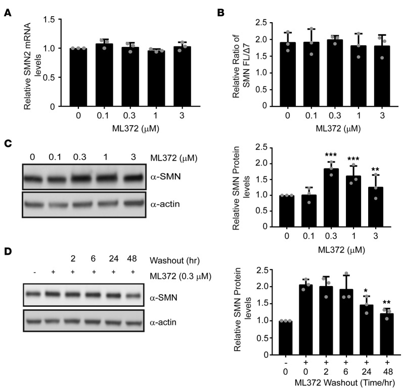
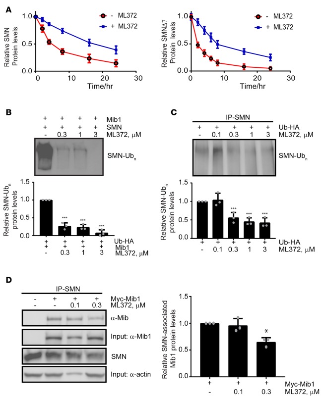
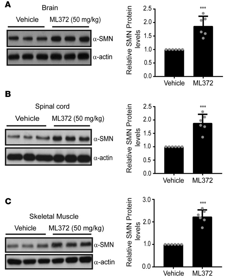
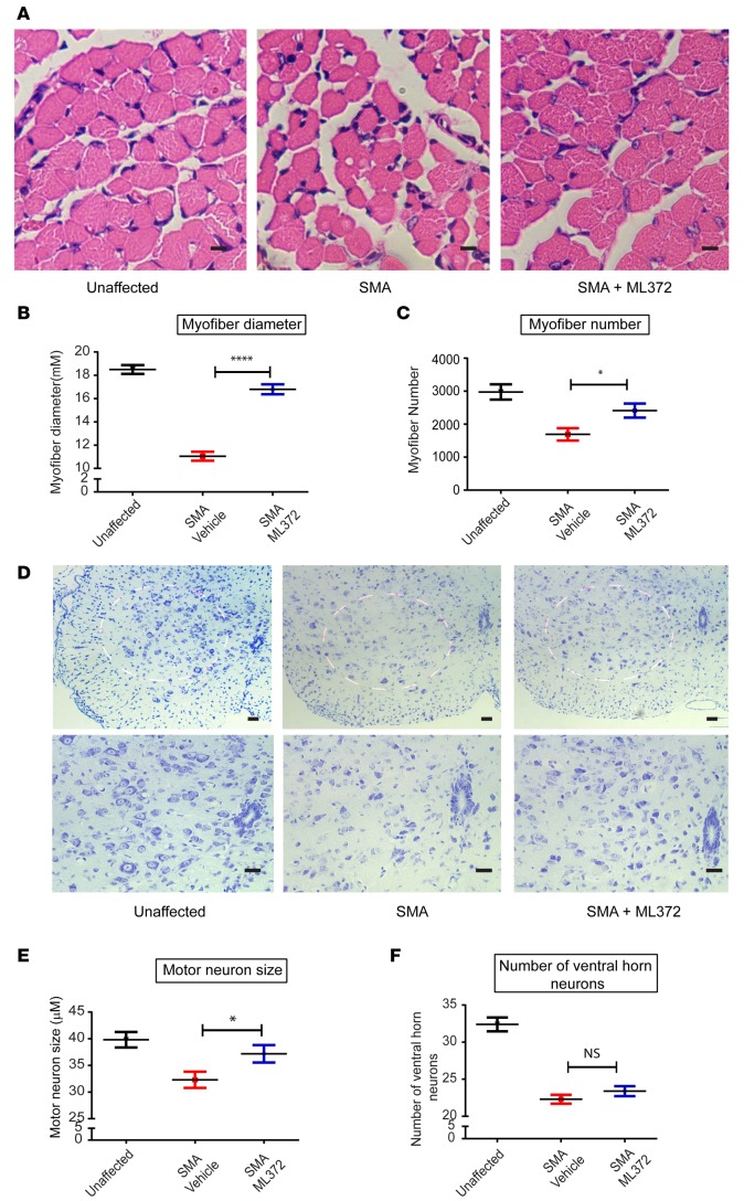
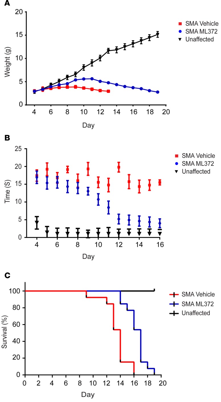
Spinal muscular atrophy (SMA) is an autosomal recessive neuromuscular disease and one of the leading inherited causes of infant mortality. SMA results from insufficient levels of the survival motor neuron (SMN) protein, and studies in animal models of the disease have shown that increasing SMN protein levels ameliorates the disease phenotype. Our group previously identified and optimized a new series of small molecules, with good potency and toxicity profiles and reasonable pharmacokinetics, that were able to increase SMN protein levels in SMA patient-derived cells. We show here that ML372, a representative of this series, almost doubles the half-life of residual SMN protein expressed from the SMN2 locus by blocking its ubiquitination and subsequent degradation by the proteasome. ML372 increased SMN protein levels in muscle, spinal cord, and brain tissue of SMA mice. Importantly, ML372 treatment improved the righting reflex and extended survival of a severe mouse model of SMA. These results demonstrate that slowing SMN degradation by selectively inhibiting its ubiquitination can improve the motor phenotype and lifespan of SMA model mice.


Conflict of interest statement

The authors declare that no conflict of interest exists.

Figures

Figure 1
Figure 1. ML372 enhances SMN protein level and function.
Spinal muscular atrophy patient fibroblast 3813 cells were treated with vehicle or ML372 at indicated concentrations for 48 hours. (A) SMN2 transcript expression was measured by qPCR. (B) The relative ratio of full-length SMN to the Δ7 mutant (FL/Δ7) was used to determine the exon 7 inclusion in SMN2 mRNA. (C) Western blot was used to determined SMN protein levels (left panel). Densitometry analysis is shown as the mean ± SEM (n = 3, **P < 0.01, ***P < 0.001) (right panel). (D) A series of wash-out experiments were performed and SMN protein level was determined by Western blot. Densitometry analysis is shown as the mean ± SEM (n = 3, *P < 0.05, **P < 0.01) (right panel). All P values determined by unpaired 2-tailed Student’s t test.
Figure 2
Figure 2. SMN ubiquitination is modulated by ML372.
(A) Pulse-chase analysis of myc-SMN (left panel) and myc-SMNΔ7 (right panel) transiently expressed in HEK-293T cells in the presence and absence of 0.3 μM ML372. (B) Recombinant SMN was incubated with the ubiquitin-activating enzyme (E1), the ubiquitin-conjugating enzyme (UBCH5b), and Mib1 with or without ML372 at the indicated concentrations. Densitometry analysis is shown as the mean ± SEM (n = 3, ***P < 0.001) (bottom panel). (C) HEK-293T cells were transiently transfected with 1 μg of HA-ubiquitin. Cells were treated with various concentrations of ML372 for 48 hours and ubiquitinated SMN was immunoprecipitated. Densitometry analysis is shown as the mean ± SEM (n = 3, ***P < 0.001) (bottom panel). (D) HEK-293T cells were transiently transfected with 1 μg of myc-Mib1. Cells were treated with various concentrations of ML372 for 48 hours, and SMN was immunoprecipitated. Western blot was used to determine SMN-associated Mib1. Densitometry analysis is shown as the mean ± SEM (n = 3, *P < 0.05) (right panel). P values were determined by 1-way ANOVA followed by Dunnett’s multiple comparisons test.
Figure 3
Figure 3. ML372 increases SMN protein levels in SMNΔ7 spinal muscular atrophy (SMA) mice.
Protein lysates of (A) brain, (B) spinal cord, and (C) muscle tissues (50–100 μg) from vehicle- or ML372-treated SMNΔ7 SMA mice were resolved by 10% SDS-PAGE. Densitometry analysis is shown as the mean ± SEM (n = 6, ***P < 0.001). P values were determined by unpaired 2-tailed Student’s t test.
Figure 4
Figure 4. ML372 increases myofiber size and number and augments ventral horn neuron size.
SMNΔ7 spinal muscular atrophy (SMA) mice were treated with vehicle or ML372 from postnatal day 5 (PND5) to PND9. Black, unaffected; Red, SMNΔ7 SMA mice treated with vehicle; Blue, SMNΔ7 SMA mice treated with ML372. (A) H&E staining of tibialis anterior (TA) muscles from unaffected, SMA, and drug-treated SMA mice. Scale bars: 100 μm. (B) Mean myofiber diameter was compared in SMA and ML372-treated SMA mice. The results are indicated as the mean ± SEM (n = 20, ****P < 0.0001). (C) Myofiber numbers were counted in the 3 groups. The analysis is shown as the mean ± SEM (n = 20, *P < 0.05). (D) Nissl-stained cross-sections of spinal cord showing ventral horn neurons. Scale bars: 100 μm (upper and lower panels magnification ×10 and ×20, respectively). (E) The size of motor neurons was analyzed in the 3 groups and the mean ventral horn neuron size was compared in SMA mice treated with vehicle or ML372. The data are shown as the mean ± SEM (n = 20, *P < 0.05). (F) The number of motor neurons was determined in the 3 groups, and the analysis is shown as the mean ± SEM (n = 20). P values were determined by unpaired 2-tailed Student’s t test.
Figure 5
Figure 5. ML372 rescues motor function and improves life span.
Blinded study was performed in litters that were randomized into vehicle- and compound-treated groups. Red, SMNΔ7 spinal muscular atrophy (SMA) mice treated with vehicle; Blue, SMNΔ7 SMA mice treated with ML372. (A) SMNΔ7 SMA mice were weighed to assess changes in body weight. (B) Motor function was determined by measuring the time to right in unaffected (Black), vehicle-, or ML372-treated SMNΔ7 SMA mice. (C) Kaplan-Meier survival analysis of SMNΔ7 SMA mice treated with vehicle or ML372; P < 0.003, log-rank test.

References

    1. Sugarman EA, et al. Pan-ethnic carrier screening and prenatal diagnosis for spinal muscular atrophy: clinical laboratory analysis of >72,400 specimens. Eur J Hum Genet. 2012;20(1):27–32. doi: 10.1038/ejhg.2011.134. - DOI - PMC - PubMed
    1. Awano T, Kim JK, Monani UR. Spinal muscular atrophy: journeying from bench to bedside. Neurotherapeutics. 2014;11(4):786–795. doi: 10.1007/s13311-014-0293-y. - DOI - PMC - PubMed
    1. D’Amico A, Mercuri E, Tiziano FD, Bertini E. Spinal muscular atrophy. Orphanet J Rare Dis. 2011;6:71. - PMC - PubMed
    1. Arnold WD, Kassar D, Kissel JT. Spinal muscular atrophy: diagnosis and management in a new therapeutic era. Muscle Nerve. 2015;51(2):157–167. doi: 10.1002/mus.24497. - DOI - PMC - PubMed
    1. Lefebvre S, et al. Identification and characterization of a spinal muscular atrophy-determining gene. Cell. 1995;80(1):155–165. doi: 10.1016/0092-8674(95)90460-3. - DOI - PubMed

Publication types

Substances