. 2016 Nov 24:7:13595.
doi: 10.1038/ncomms13595.

Repression of RNA polymerase by the archaeo-viral regulator ORF145/RIP

Affiliations

Repression of RNA polymerase by the archaeo-viral regulator ORF145/RIP

Carol Sheppard et al. Nat Commun. .

Abstract

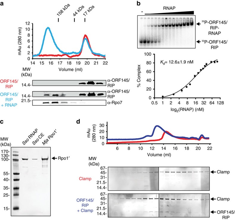
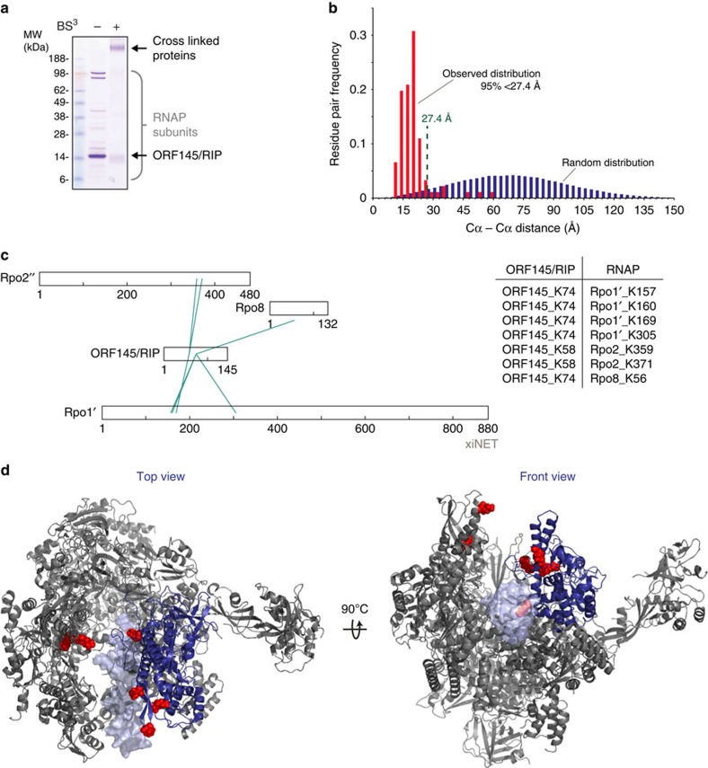
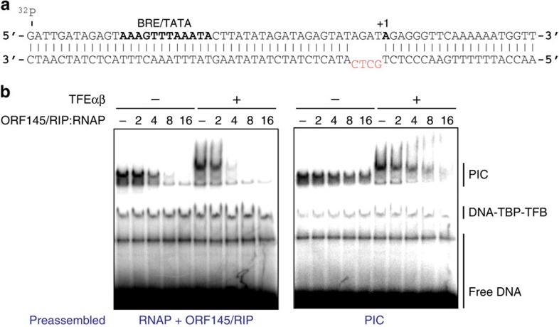
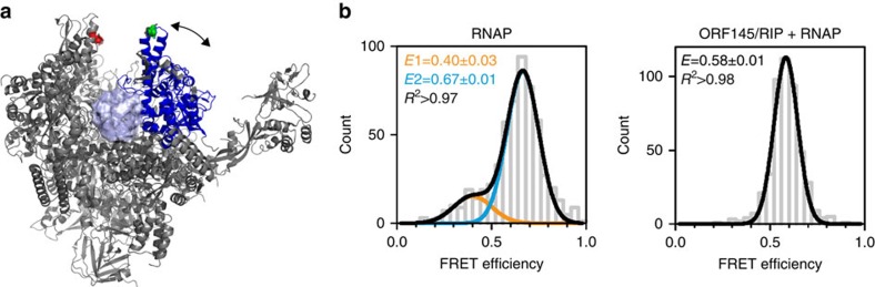
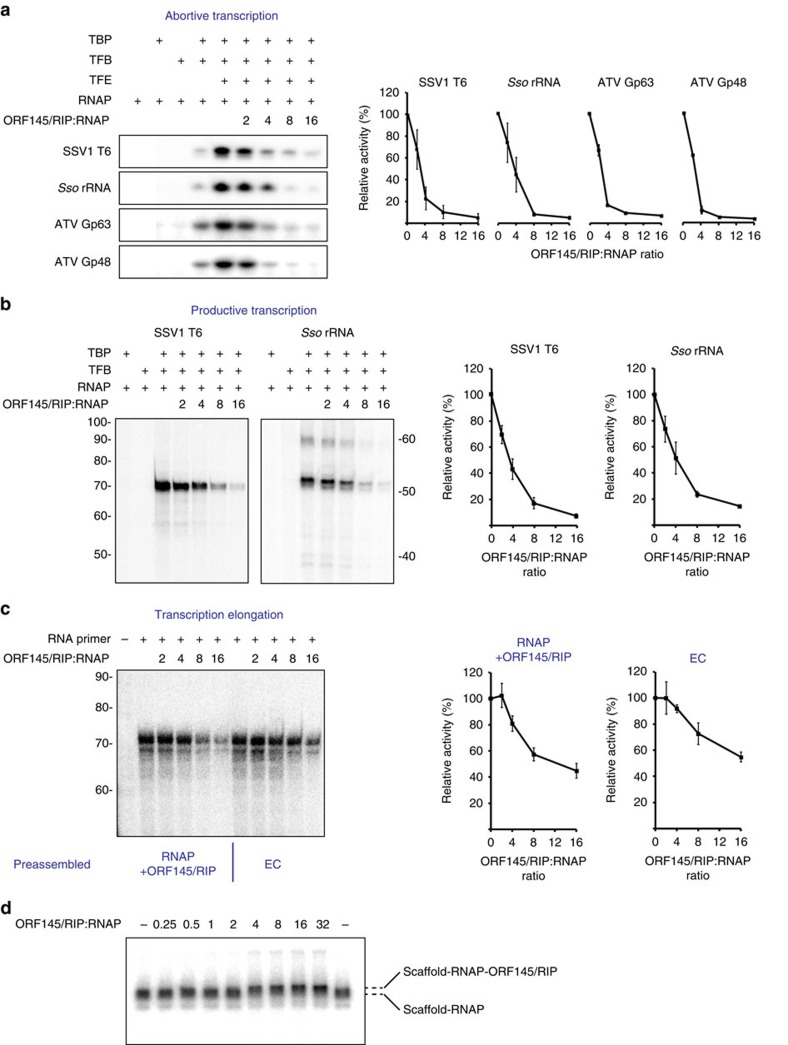
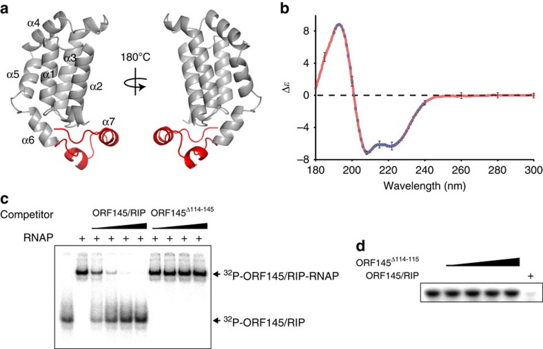
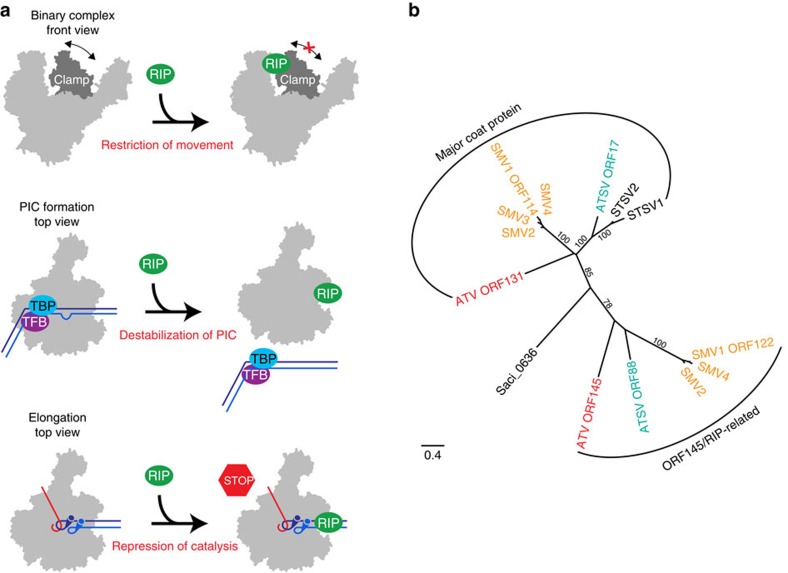
Little is known about how archaeal viruses perturb the transcription machinery of their hosts. Here we provide the first example of an archaeo-viral transcription factor that directly targets the host RNA polymerase (RNAP) and efficiently represses its activity. ORF145 from the temperate Acidianus two-tailed virus (ATV) forms a high-affinity complex with RNAP by binding inside the DNA-binding channel where it locks the flexible RNAP clamp in one position. This counteracts the formation of transcription pre-initiation complexes in vitro and represses abortive and productive transcription initiation, as well as elongation. Both host and viral promoters are subjected to ORF145 repression. Thus, ORF145 has the properties of a global transcription repressor and its overexpression is toxic for Sulfolobus. On the basis of its properties, we have re-named ORF145 RNAP Inhibitory Protein (RIP).


Figures

Figure 1
Figure 1. ORF145/RIP binds to the Sso RNAP via the Rpo1′ clamp.
(a) SEC of ORF145/RIP and ORF145/RIP–RNAP complexes. Ultraviolet elution profiles of ORF145/RIP (red trace) and ORF145/RIP pre-incubated with Sso RNAP (blue trace). Western blot analysis of SEC peak fractions detecting ‘free' ORF145/RIP (elution volume 19–21 ml), and co-elution of ORF145/RIP with Sso RNAP (elution volumes 15–17 ml). (b) EMSA of the ORF145/RIP–RNAP complex using radiolabelled ORF145/RIP. The gel bands were quantified and then plotted with the tight binding equation (see Methods for further details) from which a Kd of 12.6±1.9 nM was calculated. (c) A far-western blot was conducted by immobilizing purified RNAP and cell extract samples, probing first with recombinant ORF145/RIP protein followed by anti-ORF145/RIP polyclonal serum and finally anti-rabbit secondary antibody. ORF145/RIP interacts with the Rpo1′ subunit from the purified 13-subunit Sso RNAP, Sso cell extract, as well as purified recombinant Mja Rpo1′ subunit. (d) SEC analysis of the recombinant RNAP clamp domain (red trace) and the ORF145/RIP–clamp complex (blue trace). Error bars represent standard deviation from three technical repeats.
Figure 2
Figure 2. ORF145/RIP binds in the DNA-binding channel of RNAP.
(a) Chemical crosslinking of ORF145/RIP to the Sso RNAP. SDS–PAGE of Sso RNAP and ORF145/RIP before and after BS3 treatment. (b) Distance distribution of Lys–Lys links mapped on the RNAP structure (red bars) compared with a random distribution (blue bars) reveals that >95% of links are below the limit of the BS3 (Cα–Cα 27.4 Å). (c) Crosslinking network between RNAP and ORF145/RIP using XiNet. The table summarizes the amino-acid residues of ORF145/RIP and RNAP subunits that are crosslinked by BS3. (d) BS3-reactive lysine residues are shown on the structure of the Sso RNAP–DNA complex (pdb 4B1O, front and top views), crosslinked lysine residues are highlighted as red spheres, the RNAP clamp in blue and the double-stranded DNA template as light blue.
Figure 3
Figure 3. ORF145/RIP inhibits PIC formation.
(a) Sequence of the SSV1 T6 promoter used in PIC formation. The BRE/TATA motif and the +1 position are highlighted in bold and the non-complementary −4 to −1 regions are shown in red. (b) EMSA monitoring the formation of the archaeal PICs. Minimal archaeal PICs contain promoter DNA, TBP, TFB and RNAP. The addition of ORF145/RIP inhibits the formation of PICs in a dose–response dependent manner. The inclusion of TFEαβ leads to a supershift of the minimal PIC, which remains sensitive to ORF145/RIP. In the left panel RNAP and ORF145/RIP were incubated prior to the addition to DNA–TBP–TFB, while in the right panel PICs were allowed to form prior to the addition of ORF145/RIP.
Figure 4
Figure 4. Single-molecule FRET analysis reveals that ORF145/RIP locks the RNAP clamp in a defined conformation.
(a) RNAPs were double-labelled with a FRET donor–acceptor dye pair (DyLight500-Dylight650) at amino-acid residues Rpo1′–E257 and Rpo2′′–Q373 indicated as green and red spheres, respectively (pdb 4B1O, the mobile clamp is shown in blue). (b) Single-molecule FRET histograms of RNAP (left) and ORF145/RIP–RNAP complexes (right). The RNAP clamp exists in open and closed conformations represented as a low (E=0.40±0.03, fit with a Gaussian function shown in orange) and high (E=0.67±0.01, fit with a Gaussian shown in blue) FRET population, respectively. In contrast, the ORF145/RIP–RNAP complex exhibits a single FRET distribution with an intermediate conformation (E=0.58±0.01) that was fitted with a single Gaussian function. The mean FRET efficiencies (E) and the coefficient of determination (R2) are given with s.e.'s in the histograms.
Figure 5
Figure 5. ORF145/RIP is a potent inhibitor of transcription.
(a) Abortive initiation assay using the SSV1 T6 promoter, Sso rRNA promoter, ATV Gp63 and ATV Gp48 promoters. The addition of ORF145/RIP represses the synthesis of a trinucleotide product in a dose-dependent manner. (b) Promoter-directed transcription initiation from all promoters is strictly dependent on TBP and TFB; ORF145/RIP efficiently represses transcription from both promoters in a dose–response manner. Sso RNAP can utilize synthetic elongation scaffolds containing DNA template- and non-template strands and a short RNA primer. Transcription elongation is repressed by ORF145/RIP in a dose–response manner (c). The preassembled RNAP–DNA–RNA complex is slightly less sensitive to ORF145/RIP repression when compared with reactions where the ORF145/RIP–RNAP complex was allowed to form prior to the addition of DNA–RNA scaffold (c). In each assay the transcripts were quantified using phosphorimaging and plotted as a function of ORF145/RIP–RNAP stoichiometry (right hand graphs on panels ac). (d) EMSA demonstrating the simultaneous binding of the DNA–RNA scaffold and ORF145/RIP to RNAP. The small but significant difference in mobility between the RNAP–scaffold and the RNAP–scaffold–ORF145/RIP complexes is indicated with dashed lines. Error bars represent standard deviation from three technical repeats.
Figure 6
Figure 6. Structural homology model of ORF145/RIP.
(a) Homology model of ORF145/RIP prepared with I-TASSER (C-score −2.12, TM-score 0.46±0.15) shown as a cartoon representation with the C-terminal tail (114–145) coloured red. (b) CD spectrum of ORF145/RIP (red trace) confirms a very high alpha-helical content of the recombinant protein when compared with the spectra of proteins with known structures in Dichroweb (blue circles). (c) Truncation of the small C-terminal tail (Δ114–145) abrogates the binding of ORF145/RIP to RNAP. The EMSA shows that unlabelled wild-type ORF145/RIP competes for 32P-ORF145/RIP binding to RNAP, while ORF145Δ114–145 does not. (d) ORF145Δ114–145 does not inhibit abortive transcription. Error bars represent standard deviation of at least eight separate spectra.
Figure 7
Figure 7. Molecular mechanism and evolution of ORF145/RIP.
(a) Schematic illustration of the molecular mechanism of RNAP repression. ORF145/RIP binds in the DNA-binding channel of RNAP and restricts the movement of the RNAP clamp. This destabilizes PIC complexes, and thus inhibits abortive and productive transcription. ORF145/RIP binds to RNAP simultaneously with the DNA/RNA template resulting in a repressed form of the transcription elongation complex. (b) The ORF145/RIP phylogenetic tree; maximum likelihood phylogenetic reconstruction of ATV ORF145/RIP and ORF131 and related proteins. Phylogenetic analysis suggests that the viral proteins fall into two different clades: ORF145/RIP-related and ORF131-related (major coat proteins). The scale bar represents amino-acid changes per site. Bootstrap values (100 replicates) are shown next to each branch. An alignment of ATV_ORF131 and ORF145/RIP-related proteins is available in Supplementary Fig. 5.

Similar articles

Cited by

References

    1. Gottesman M. Bacteriophage lambda: the untold story. J. Mol. Biol. 293, 177–180 (1999). - PubMed
    1. Gottesman M. E. & Weisberg R. A. Little lambda, who made thee? Microbiol. Mol. Biol. Rev. 68, 796–813 (2004). - PMC - PubMed
    1. Forterre P. & Prangishvili D. The major role of viruses in cellular evolution: facts and hypotheses. Curr. Opin. Virol. 3, 558–565 (2013). - PubMed
    1. Prangishvili D., Forterre P. & Garrett R. A. Viruses of the Archaea: a unifying view. Nat. Rev. Microbiol. 4, 837–848 (2006). - PubMed
    1. Ortmann A. C., Wiedenheft B., Douglas T. & Young M. Hot crenarchaeal viruses reveal deep evolutionary connections. Nat. Rev. Microbiol. 4, 520–528 (2006). - PubMed

Publication types

MeSH terms

Substances