. 2016 Nov 24:6:37708.
doi: 10.1038/srep37708.

Structural and functional probing of PorZ, an essential bacterial surface component of the type-IX secretion system of human oral-microbiomic Porphyromonas gingivalis

Affiliations

Structural and functional probing of PorZ, an essential bacterial surface component of the type-IX secretion system of human oral-microbiomic Porphyromonas gingivalis

Anna M Lasica et al. Sci Rep. .

Abstract

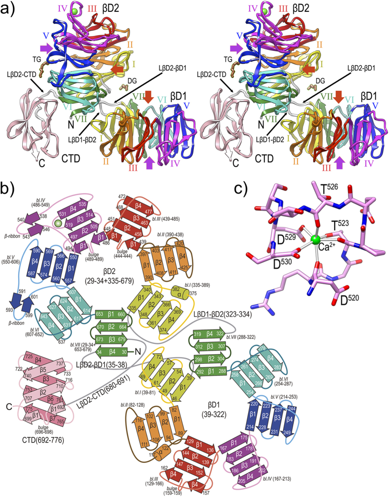
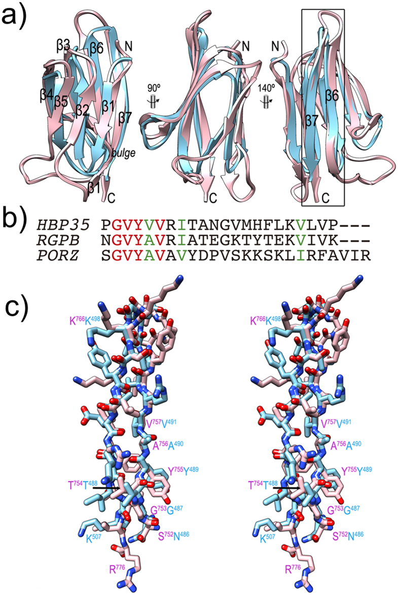
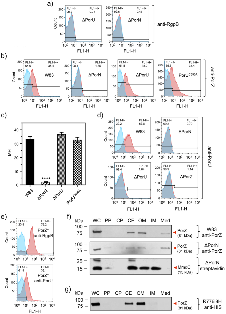
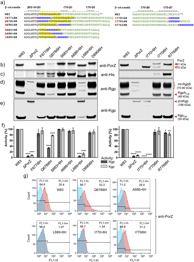
Porphyromonas gingivalis is a member of the human oral microbiome abundant in dysbiosis and implicated in the pathogenesis of periodontal (gum) disease. It employs a newly described type-IX secretion system (T9SS) for secretion of virulence factors. Cargo proteins destined for secretion through T9SS carry a recognition signal in the conserved C-terminal domain (CTD), which is removed by sortase PorU during translocation. Here, we identified a novel component of T9SS, PorZ, which is essential for surface exposure of PorU and posttranslational modification of T9SS cargo proteins. These include maturation of enzyme precursors, CTD removal and attachment of anionic lipopolysaccharide for anchorage in the outer membrane. The crystal structure of PorZ revealed two β-propeller domains and a C-terminal β-sandwich domain, which conforms to the canonical CTD architecture. We further documented that PorZ is itself transported to the cell surface via T9SS as a full-length protein with its CTD intact, independently of the presence or activity of PorU. Taken together, our results shed light on the architecture and possible function of a novel component of the T9SS. Knowledge of how T9SS operates will contribute to our understanding of protein secretion as part of host-microbiome interactions by dysbiotic members of the human oral cavity.


Figures

Figure 1
Figure 1. Characterization of the ΔPorZ secretory phenotype.
(a) Pigmentation on blood agar of P. gingivalis wild type (W83), ΔPorZ, and in trans porZ-complemented ΔPorZ (PorZ+) strains. Enzymatic activity of (b) Rgps, (c) Kgp, and (d) dipeptidyl peptidase IV (DPPIV) and prolyl tripeptidyl peptidase (PtpA) in whole cultures, fractionated washed cells or growth medium as determined with specific synthetic substrates. Cultures were adjusted to OD600 = 1.0 prior to testing and processing, and results shown correspond to triplicate experiments. Significant differences between the wild type and mutants are indicated by *P < 0.05 and ****P < 0.0001.
Figure 2
Figure 2. Subcellular location of gingipains and PPAD.
Whole cells (WC) of wild-type (W83; left panel) and ΔPorZ (right panel) P. gingivalis strains were proportionately fractionated into periplasm (PP), cytoplasm (CP), cell envelope (CE), outer membrane (OM), inner membrane (IM) and culture medium fractions (Med; 10-fold concentrated); and probed for (a) Rgps, (b) Kgp, (c) PPAD by Western blotting with specific monoclonal antibodies and (d) biotinylated IM protein (MmdC) through reaction with streptavidin conjugated to horseradish peroxidase. The pinpointed and labeled bands correspond to: (a) catalytic domain of RgpA (RgpAcat) and membrane-type RgpB (mt-RgpB) in the wild type (left panel) and unprocessed pro-RgpA and pro-RgpB in ΔPorZ (right panel); (b) catalytic domain of Kgp (Kgpcat) in the wild type (left panel) and unprocessed pro-Kgp in ΔPorZ (right panel); (c) mature PPAD and membrane-type PPAD (mt-PPAD) in the wild type (left panel) and unprocessed pro-PPAD in ΔPorZ (right panel); and (d) MmdC in the wild-type (left panel) and ΔPorZ (right panel) strains.
Figure 3
Figure 3. PorZ is located on the cell surface of P. gingivalis.
(a) Wild-type P. gingivalis cells (W83) were proportionately fractionated into whole cell extract (WC), periplasm (PP), cytoplasm (CP), cell envelope (CE), outer membrane (OM), inner membrane (IM) and growth medium (Med), and subsequently analyzed by Western blotting using mouse polyclonal anti-PorZ and anti-Rgp antibodies and mouse monoclonal anti-Kgp antibodies. Streptavidin conjugated to horseradish peroxidase was used to detect MmdC, a biotinylated IM-associated protein. Presence or absence of full-length PorZ (81-kDa band) and other proteins is indicated. (b) Wild-type cells were probed with monoclonal anti-PorZ antibodies and labeled with immunogold to visualize the cellular location of PorZ (black arrowhead) in electron microscopy (bar = 100 nm). (c) Dot blot analysis of intact and lyzed wild-type (W83), ΔPorZ, ΔPorU and ΔPorN cells using mouse monoclonal anti-PorZ antibodies (mAb), mouse polyclonal anti-PorZ antibodies (pAb) or streptavidin conjugated to horseradish peroxidase. Flow cytometry analysis showing the surface exposure of (d) PorZ in wild-type cells (W83) with anti-PorZ pAb; (e) RgpB in wild-type cells (W83) with monoclonal anti-RgpB antibodies (positive control); (f) MmdC in wild-type cells (W83) and (g) in ΔPorZ cells with streptavidin-Alexa Fluor 488 conjugate; (h) PorZ in in trans porZ-complemented ΔPorZ (PorZ+) cells with pAb and (i) PorZ in ΔPorZ cells with anti-PorZ pAb. Isotype negative controls are in blue and immunoprobed cells in red; the histograms shown are representative of three independent experiments. (j) Presence of full-length PorZ (lane 1) detected by Western blot using anti-PorZ pAb on wild-type cells washed with PBS and suspended, respectively, in distilled water (lane2); in 0.0007% Tween-20 (lane 3); in 0.04% sarcosyl (lane 4); and in 0.02% SDS (lane 5). After 10 min of gentle stirring, cells were removed by centrifugation and the presence of PorZ in the cell pellet (lane 1) and supernatants (lanes 2–5 was checked). The detergent concentrations correspond to one-tenth of the critical micelle concentration (CMC). Lane M, MagicMark™ XP Western Protein Standard.
Figure 4
Figure 4. Effect of PorZ on the expression of T9SS components and cargos.
The respective mRNAs of the genes of T9SS components (porT, sov, porU, lptO, porN, porO and porW) and cargos (rgpB, kgp and cpg70) were quantified in mid-logarithmic cultures of wild type (W83; full bars) and ΔPorZ (open bars) cells by quantitative RT-PCR. Expression levels were normalized against ribosomal r16s expression, and the respective expression levels of the wild type were arbitrarily set to 1.0. The results correspond to four independent experiments. Error bars represent standard deviation as analyzed by Student’s t-test. *P < 0.05; **P < 0.01 and ***P < 0.001.
Figure 5
Figure 5. Overall crystal structure of PorZ.
(a) Ribbon-type plot in cross-eye stereo of the crystal structure to 2.9 Å resolution of PorZ depicting domains βD1, βD2 and CTD, and the three domain-connecting linkers (white ribbons; labeled LβD2-βD1, LβD1-βD2, and LβD2-CTD). Each of the seven blades of propellers βD1 and βD2 (labeled counter-clockwise I to VII) is colored in yellow, orange, red, magenta, blue, turquoise and green, respectively; the CTD is in pink. A structural calcium-binding site (green sphere) is found within βD1-blade IV, and a tetraethylene glycol (TG) and a diethylene glycol (DG) were tentaively assigned on the protein surface (brown stick-models). Other (functionally probably irrelevant) ions and ligands were omitted for clarity. The central shafts of βD1 and βD2 are pinpointed on the entry and exit sides of the propellers by red and purple arrows, respectively. For labels and extension of regular secondary structure elements, see (b). (b) Topology scheme of PorZ, with β-strands as arrows and helices as cylinders, colored as in (a). The polypeptide chain spans residues G29—R776 and the three constituting domains plus the linkers (in grey) are indicated with the residues delimiting each structural element (strands, bulges, helices, β-ribbons, blades and domains). The nomenclature adopted in the text for structure elements is “domain-blade-structural element”, e.g. βD1-VI-β3 or βD2-IV-β-ribbon. (c) Structural calcium-binding site framed by segment D520—D530 within loop Lβ3β4 of βD1-blade IV. The ion is octahedrally coordinated by D520O, T523O, T523Oγ, T526O, D529Oδ1 and D530Oδ1, which are at binding distances of ~2.4 Å.
Figure 6
Figure 6. The C-terminal domain of PorZ.
(a) Ribbon-plot of the CTDs of PorZ (pink) and RgpB (cyan) after optimal superposition facing the three-stranded front β-sheet (left, the seven constituting strands are labelled, see also Fig. 5b); after a vertical 90° rotation (center); and facing the four-stranded back β-sheet (right). C-terminal strands β6 and β7, which contain the reported molecular determinants for T9SS secretion, are framed and labeled in the right panel. (b) Sequence alignment of the 22 C-terminal residues of HBP35 (UniProt Q8G962) and RgpB (UniProt P95493), and the 25 final residues of PorZ after the structural alignment of the structures of the latter two proteins (see also [c]). Identical residues are in red, similar ones in green. G-X-Y sequences are also found in PKD proteins within strands equivalent to CTD-β6. These are also seven-stranded immunoglobulin-like all-β domains, although the function of the G-X-Y motif therein is unknown. In addition, the Y_Y_Y domain of BT4663 protein contains this signature. (c) Detail in cross-eye stereo showing the strands framed in (a) as full-atom models, i.e. segments S752—R776 of PorZ (with pink carbons and magenta labels) and N486—K507 of RgpB (mature protein numbering as subscripts, add 229 for full-length protein numbering; cyan carbons and labels).
Figure 7
Figure 7. PorZ is secreted via T9SS with an intact CTD independently of PorU.
Flow cytometry analysis using (a) anti-RgpB mAbs in ΔPorU and ΔPorN; (b) anti-PorZ antibodies in the wild type (W83), PorN-null mutant (ΔPorN), PorU-null mutant (ΔPorU), and PorU active-site inactivation mutant (PorUC690A); (c) PorZ surface exposure as mean fluorescent intensity (MFI) in different strains calculated from flow cytometry analysis (in duplicates) from three different cultures. Significant differences between the wild type and mutants were analyzed by one-way ANOVA with Bonferroni’s correction; ****P < 0.0001. Flow cytometry analysis using (d) anti-PorU antibodies in the wild type (W83), PorN-null mutant (ΔPorN), PorU-null mutant (ΔPorU), and ΔPorZ; (e) anti-RgpB and anti-PorU in porZ complemented strain (PorZ+). The result of using specific antibodies (red surface) and the negative isotype control (blue surface) are shown. (f) Western blot analysis of subcellular locations of PorZ in the ΔPorN mutant by probing with anti-PorZ antibodies as compared to the wild type (W83). Streptavidin conjugated to horseradish peroxidase was used to detect MmdC, a biotinylated IM-associated control protein. (g) Same as (f) but using anti-His antibodies to detect the CTD of PorZ in the strain expressing PorZ with an octahistidine tag at the C-terminus (R776i8H). Bacterial cultures were fractionated as described in the Methods section.
Figure 8
Figure 8. Effects of the introduction of, or replacement with, oligohistidines on PorZ function.
(a) Location of insertions (i) and substitutions (>) of consecutive residues by polyhistidines at the junction (residues G680-G691, over yellow background) between CTD (green font) and the preceding domain of PorZ (F677i8H, Q678i6H, S683 > 6 H, A686 > 6 H, L689 > 6 H and D690i6H) or at the C-terminus (I770 > 6 H, I770i6H, and R776i8H). β-strands are indicated above the alignment. (be) Wild-type and mutant strains were grown to OD600 = 1.0 and whole cultures were subjected to Western blot analysis with anti-PorZ (b), anti-polyhistidine (c), anti-Rgp (d) and anti-Kgp (e) antibodies. (f) The same strains were used for gingipain activity assays. (g) The level of surface exposure of PorZ in various mutants was analyzed by flow cytometry using anti-PorZ antibodies (red) and negative isotype control (blue). Representative histograms are shown from three independent experiments.

Similar articles

Cited by

References

    1. Zückert W. R. Secretion of bacterial lipoproteins: through the cytoplasmic membrane, the periplasm and beyond. Biochim. Biophys. Acta 1843, 1509–1516 ( 2014). - PMC - PubMed
    1. Abby S. S. et al. Identification of protein secretion systems in bacterial genomes. Sci. Rep. 6, 23080 (2016). - PMC - PubMed
    1. Desvaux M., Hebraud M., Talon R. & Henderson I. R. Outer membrane translocation: numerical protein secretion nomenclature in question in mycobacteria. Trends Microbiol. 17, 338–340 (2009). - PubMed
    1. Sato K. et al. A protein secretion system linked to bacteroidete gliding motility and pathogenesis. Proc. Natl. Acad. Sci. USA 107, 276–281 (2010). - PMC - PubMed
    1. McBride M. J. & Zhu Y. Gliding motility and Por secretion system genes are widespread among members of the phylum Bacteroidetes. J. Bacteriol. 195, 270–278 (2013). - PMC - PubMed

Publication types

MeSH terms

LinkOut - more resources