. 2016 Nov 24:6:37887.
doi: 10.1038/srep37887.

Non-RVD mutations that enhance the dynamics of the TAL repeat array along the superhelical axis improve TALEN genome editing efficacy

Affiliations

Non-RVD mutations that enhance the dynamics of the TAL repeat array along the superhelical axis improve TALEN genome editing efficacy

Naoya Tochio et al. Sci Rep. .

Abstract

Transcription activator-like effector (TALE) nuclease (TALEN) is widely used as a tool in genome editing. The DNA binding part of TALEN consists of a tandem array of TAL-repeats that form a right-handed superhelix. Each TAL-repeat recognises a specific base by the repeat variable diresidue (RVD) at positions 12 and 13. TALEN comprising the TAL-repeats with periodic mutations to residues at positions 4 and 32 (non-RVD sites) in each repeat (VT-TALE) exhibits increased efficacy in genome editing compared with a counterpart without the mutations (CT-TALE). The molecular basis for the elevated efficacy is unknown. In this report, comparison of the physicochemical properties between CT- and VT-TALEs revealed that VT-TALE has a larger amplitude motion along the superhelical axis (superhelical motion) compared with CT-TALE. The greater superhelical motion in VT-TALE enabled more TAL-repeats to engage in the target sequence recognition compared with CT-TALE. The extended sequence recognition by the TAL-repeats improves site specificity with limiting the spatial distribution of FokI domains to facilitate their dimerization at the desired site. Molecular dynamics simulations revealed that the non-RVD mutations alter inter-repeat hydrogen bonding to amplify the superhelical motion of VT-TALE. The TALEN activity is associated with the inter-repeat hydrogen bonding among the TAL repeats.


Figures
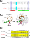
Figure 1
Figure 1. TALE sequence and structure.
(a) Comparison between the amino acid sequences of the first 4 TAL-repeats of TALE with/without the periodically mutated repeats. The sequences of TAL-repeats without (CT-TALE, upper) and with the periodical mutations (VT-TALE, lower) are presented. RVDs are shaded in cyan. Non-RVD residues at the 4th and 32nd positions are presented as bold letters with different colours for the mutation sites. The secondary structures are presented at the bottom. (b) Ribbon representations of a single-TAL repeat (left) and the DNA bound TALE protein (right) (PDBID: 3V6T14). The first short helix (helix a) and the second long helix (helix b) are coloured red and green, respectively. RVD and the periodical mutation sites are noted in cyan and magenta, respectively, with the side chains shown as a stick representation. In the right panel, RVDs and the mutation sites are also displayed. DNA is also presented in orange. Figures were prepared using PyMOL (DeLano Scientific, San Carlos, CA). (c) Schematic representation of CT-TALE (upper) and VT-TALE (lower). TAL-repeats are represented as yellow boxes. Non-canonical pseudo-repeats and the last half repeats are presented as dashed boxes and white boxes, respectively. Amino acid types of non-RVD residues at the 4th and 32nd positions in each repeat are given as letters; mutated sites are coloured.
Figure 2
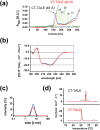
Figure 2. Comparison of physicochemical properties between CT-TALE and VT-TALE.
(a) Analyses of size exclusion chromatograms of TALEs. Calibration of the column was performed using a standard globular protein set (GE Healthcare), and the chromatogram is presented in green. Stokes radii of the standard proteins are labelled (Å unit). The chromatograms of CT-TALE and VT-TALE are indicated in blue and red, respectively. Estimated Stokes radii of CT-TALE and VT-TALE are shown in parentheses. (b) CD spectra of TALEs. CD spectra of CT-TALE and VT-TALE are presented in blue and red, respectively. (c) Size distributions of CT- and VT-TALE. Particle size distributions of CT- and VT-TALE are presented in blue and red, respectively. Estimated Z-average sizes were 48.3 ± 0.7 Å and 44.7 ± 1.6 Å, respectively. (d) DSC thermograms of the CT- and VT-TALEs. Experimental data are shown in black, and fits for individual and composite fits are indicated in red. (upper) CT-TALE: T1m = 61.71 ± 0.02 °C; T2m = 63.41 ± 0.01 °C, (lower) VT-TALE: Tm = 52.07 ± 0.01 °C.
Figure 3
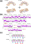
Figure 3. Inter-repeat hydrogen bonds among the TAL-repeats.
(a) Snapshots of the CT- and VT-TALE models in the extended forms sampled at 50 ns in the MD trajectories. The structures are viewed from the C-terminal side of the superhelical structure and its 90-degree rotated position. For the four TAL-repeats in the centre of the array, the side chains of the residues positioned at 4 and 5 in each repeat of CT- and VT-TALEs are drawn as ball and stick representations. Residue at position 4, a non-RVD residue, in each TAL-repeat is marked in purple. For details of the modelling and simulation, see the Supplementary Information. (b) Distances between HE21/HE22 in Q-5 and OD1/OD2 in D-4 or OE1/OE2 in E-4 in the structures in the MD trajectory (corresponding to Trial 1 in Supplementary Fig. S4); every atomic pair is considered. (c) Schematic representation of the inter-repeat hydrogen bonds. Helices a and b and RVD are presented as magenta boxes, green boxes and cyan lines, respectively. The 4th and 5th amino acids in each TAL-repeat are represented by short lines. Hydrogen bonds are indicated by orange lines. In VT-TALE, two of the inter-repeat hydrogen bonds are not formed in a unit with four TAL-repeats.
Figure 4
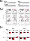
Figure 4. Comparison between the DNA binding mechanism of VT-TALE and CT-TALE.
(a) The DNA sequences used in this study. EBEf (effector binding element): full target sequence; non-EBE: non-target sequence; EBEn: N-terminal half (5′ side) TAL-repeat array target sequence; EBEc: C-terminal half (3′ side) TAL-repeat array target sequence. The underlined nucleotides are target sequences recognised by our TALE samples. (b) ITC analyses of CT-TALE and VT-TALE against a series of DNAs. Each plot of the total heat released is presented as a function of the molar ratio of added DNA, which was subtracted by the heat generated owing to the dilution of the titrants. The red lines represent the nonlinear, least-squares best fit to the experimental data, using a one-site model. (c) The thermodynamic signatures of TALEs binding to EBEf (left) and EBEn (right). The upper and lower panels indicate the results at 15 and 25 °C, respectively. The results of CT-TALE and VT-TALE are presented in black and red, respectively. All experiments were carried out twice.
Figure 5
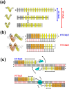
Figure 5. Enhanced superhelical motion by the non-RVD mutations to TALEN.
(a) The superhelical amplitude motion of VT-TALE is greater in magnitude compared with that of CT-TALE. The model TALE structures in different conformations occurred in the molecular dynamics simulation are located on the left to the schematic drawings. The canonical TAL-repeats, N-terminal atypical repeats and C-terminal half repeats are indicated as yellow, grey and white boxes, respectively. (b) The number of TAL-repeats in VT-TALE (lower) that engage in DNA binding is greater than that observed for CT-TALE (upper). The DNA-bound TAL-repeats are represented by orange boxes, and the free TAL-repeats are drawn as yellow boxes. The DNA bases specifically recognised by the TAL-repeats are presented as red bars. The modelled TALE structures in the complex with DNA are located on the left of the schematic drawings for the complexes having a different number of TAL-repeats engaged in the DNA binding. (c) The spatial restriction of the FokI domains by the extended TAL-repeat interaction to DNA in VT-TALE (lower) could facilitate FokI dimerisation more readily than that observed for CT-TALE (upper). FokI monomers are depicted as cyan semicircles. The arrows schematically represent the magnitudes of the structural dynamics of the TAL-repeats not bound to DNA and the FokI domain associated with the C-terminal end of the TAL-repeat array.

Similar articles

Cited by

References

    1. Gu K. et al.. R gene expression induced by a type-III effector triggers disease resistance in rice. Nature 435, 1122–1125 (2005). - PubMed
    1. Kay S., Hahn S., Marois E., Hause G. & Bonas U. A Bacterial Effector Acts as a Plant Transcription Factor and Induces a Cell Size Regulator. Science 318, 648–651 (2007). - PubMed
    1. Romer P. et al.. Plant Pathogen Recognition Mediated by Promoter Activation of the Pepper Bs3 Resistance Gene. Science 318, 645–648 (2007). - PubMed
    1. White F. F. & Yang B. Host and Pathogen Factors Controlling the Rice-Xanthomonas oryzae Interaction. PLANT Physiol. 150, 1677–1686 (2009). - PMC - PubMed
    1. Scholze H. & Boch J. TAL effectors are remote controls for gene activation. Curr. Opin. Microbiol. 14, 47–53 (2011). - PubMed

Publication types

MeSH terms

Substances

LinkOut - more resources