Case Reports
. 2017 Jan;31(1):142-148.
doi: 10.1111/jvim.14609. Epub 2016 Nov 24.

Pyogranulomatous Pancarditis with Intramyocardial Bartonella henselae San Antonio 2 (BhSA2) in a Dog

Affiliations
Case Reports

Pyogranulomatous Pancarditis with Intramyocardial Bartonella henselae San Antonio 2 (BhSA2) in a Dog

T A Donovan et al. J Vet Intern Med. 2017 Jan.
No abstract available

Keywords: Canine; Myocarditis; Nephritis; Stealth; Vasculitis.


Figures

Figure 1
Figure 1
Heart, dog. Myriad, multifocal to coalescing white to tan nodular foci are visible from the epicardium of the right and left ventricles and atria. Bar = 5 mm.
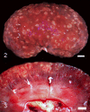
Figure 2 and 3
Figure 2 and 3
Kidney, dog. Visible from the renal cortex are dozens of multifocal to coalescing, tan nodular foci, which range from pinpoint to 1 cm in diameter. Bar = 5 mm.
Figure 4
Figure 4
Heart, dog. Low magnification view of the right ventricle and atrium. Multifocal to coalescing regions of increased cellular density (pyogranulomatous inflammation) are visible in the epicardium, myocardium, and endocardium, consistent with pancarditis. H&E. Bar = 500 μm. Inset. Higher magnification view of pyogranulomatous inflammation within the left ventricular myocardium. Neutrophils and macrophages are observed throughout the myocardial interstitium, separating cardiomyocytes. Regional cardiomyocytes undergo degeneration and necrosis (arrow), with sarcoplasm hypereosinophilia, loss of visible striations, and karyorrhectic debris. HE. Bar = 20 μm.
Figure 5
Figure 5
Kidney, dog. Low magnification view of a section of kidney. Large regions of multifocal to coalescing hypercellularity are present within the cortex, representing pyogranulomatous nephritis. HE. Bar = 500 μm. Inset. Higher magnification view of a section of kidney. Neutrophils and macrophages are observed throughout the interstitium. The renal tubular epithelial cells undergo degeneration, necrosis, and regeneration, with cytoplasmic hypereosinophilia, nuclear pyknosis, karyolysis (arrow), and crowding of tubular epithelial nuclei. HE. Bar = 20 μm.
Figure 6
Figure 6
Lung, dog. Photomicrograph of a section of lung. The interstitium is hypercellular, containing mixed inflammatory infiltrates, including neutrophils, macrophages, lymphocytes, and plasma cells. Hemorrhage, edema, and alveolar macrophages are present within the alveolar spaces. Inflammatory cells extend into the wall of an arteriole (arteritis, arrow). HE. Bar = 50 μm.
Figure 7
Figure 7
Multiphoton laser scanning microscopy demonstrates Bartonella henselae immunoreactivity (green, large white arrows) in the myocardium. Fluorescence excitation was at 970 nm, and emission data were collected at 535 nm to capture the green channel epifluorescence and 620 nm to capture red channel epifluorescence. Under these image capture settings, there is no contribution by lipofuscin in the green channel. Erythrocytes (asterisks) appear yellow due hemoglobin autofluorescence in both the red and green emission channels (535 and 609 nm). Inflammatory cells do not autofluoresce (not visible). Image is a single in‐focus projection of 22 0.45‐μm‐thick optical z‐sections captured with an Apo LWD 25X 1.10W DIC N2 objective on a Nikon A1RMP multiphoton microscope in the University Imaging Centers at the University of Minnesota. Orthogonal or right angle data, YZ and XZ projections, on the right and bottom margins of the image (respectively) display the 3‐dimensional data in a 2‐dimensional format. Scale bar = 20 μm.

Similar articles

Cited by

References

    1. Pomerance A, Whitney JC. Heart valve changes common to man and dog: A comparative study. Cardiovasc Res 1970;4:61–66. - PubMed
    1. Varanat M, Broadhurst J, Linder KE, et al. Identification of Bartonella henselae in 2 cats with pyogranulomatous myocarditis and diaphragmatic myositis. Vet Pathol 2012;49:608–611. - PubMed
    1. Balakrishnan N, Alexander K, Keene B, et al. Successful treatment of mitral valve endocarditis in a dog associated with Actinomyces canis‐like infection. J Vet Cardiol 2016;18:271–277. - PubMed
    1. Wacnik PW, Baker CM, Herron MJ, et al. Tumor‐induced mechanical hyperalgesia involves CGRP receptors and altered innervation and vascularization of DsRed2 fluorescent hindpaw tumors. Pain 2005;115:95–106. - PubMed
    1. Zipfel WR, Williams RM, Christie R, et al. Live tissue intrinsic emission microscopy using multiphoton‐excited native fluorescence and second harmonic generation. Proc Natl Acad Sci USA 2003;100:7075–7080. - PMC - PubMed

Publication types

Associated data

LinkOut - more resources