. 2017 Feb;246(2):135-147.
doi: 10.1002/dvdy.24476.

Retinoic acid signaling regulates Krt5 and Krt14 independently of stem cell markers in submandibular salivary gland epithelium

Affiliations

Retinoic acid signaling regulates Krt5 and Krt14 independently of stem cell markers in submandibular salivary gland epithelium

Timur M Abashev et al. Dev Dyn. 2017 Feb.

Abstract

Background: Retinoic acid (RA), the active metabolite of vitamin A, has been demonstrated to be important for growth and branching morphogenesis of mammalian embryonic salivary gland epithelium. However, it is not known whether RA functions directly within epithelial cells or in associated tissues that influence morphogenesis of salivary epithelium. Moreover, downstream targets of RA regulation have not been identified.

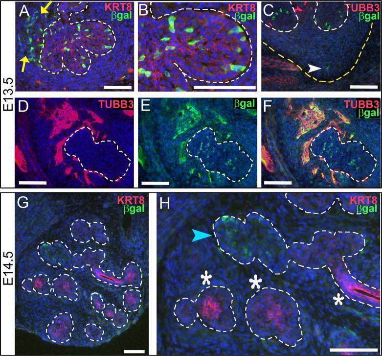
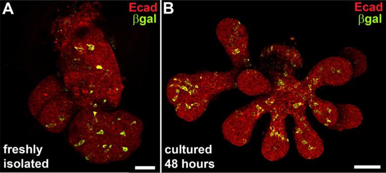
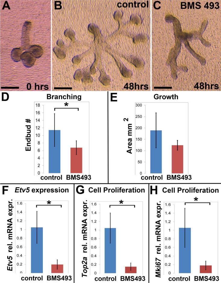
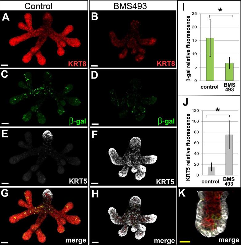
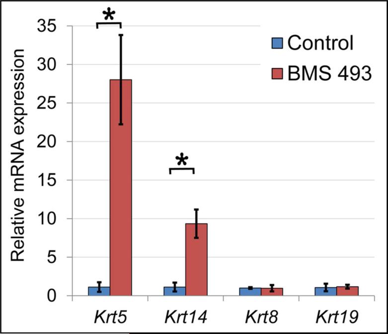
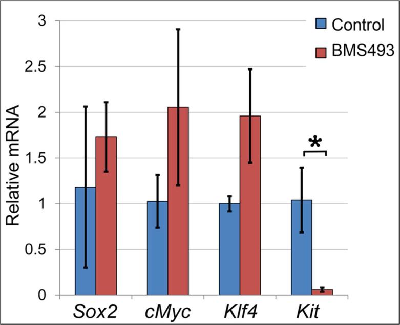
Results: Here, we show that canonical RA signaling occurs in multiple tissues of embryonic mouse salivary glands, including epithelium, associated parasympathetic ganglion neurons, and nonneuronal mesenchyme. By culturing epithelium explants in isolation from other tissues, we demonstrate that RA influences epithelium morphogenesis by direct action in that tissue. Moreover, we demonstrate that inhibition of RA signaling represses cell proliferation and expression of FGF10 signaling targets, and upregulates expression of basal epithelial keratins Krt5 and Krt14. Importantly, we show that the stem cell gene Kit is regulated inversely from Krt5/Krt14 by RA signaling.

Conclusions: RA regulates Krt5 and Krt14 expression independently of stem cell character in developing salivary epithelium. RA, or chemical inhibitors of RA signaling, could potentially be used for modulating growth and differentiation of epithelial stem cells for the purpose of re-populating damaged glands or generating bioengineered organs. Developmental Dynamics 246:135-147, 2017. © 2016 Wiley Periodicals, Inc.

Keywords: KRT5; keratin; progenitor; retinoid; stem cell; vitamin A.


Figures

Figure 1
Figure 1. RA signaling occurs in multiple tissues of developing SMG
Immunostaining for β-galactosidase on cryosections from embryos carrying the RARE-LacZ reporter transgene reveals location of RA signaling in developing SMG at E13.5 (A-F) and E14.5 (G-H). (A, B) At E13.5 co-staining for KRT8 and β-galactosidase reveals numerous cells of the epithelium are positive for RA signaling (cells within dotted outline). Cells positive for RA signaling are also detected in non-epithelial cells, particularly in the area around the main duct (yellow arrows). Examination of an individual endbud (B, (detail of A)) reveals that RA positive cells are present in basal epithelium and also within interior epithelium that will later form lumens. Co-staining for neurons (TUBB3) and RA signaling (β-galactosidase) on a posterior section through the apex of the strawberry-shaped SMG (C) reveals RA signaling occurs in a small number of cells in non-neuronal mesenchyme in this region (white arrowhead). (D-F) Co-staining for neurons (TUBB3) and RA signaling (β-galactosidase) in sections containing the parasympathetic ganglion reveals that many neurons of the SMG parasympathetic ganglion are positive for RA signaling at E13.5. (G,H) By E14.5, the number of SMG cells positive for RA signaling is reduced relative to E13.5. (G) At E14.5 RA positive cells are present in only a few endbuds. (H) KRT8 is expressed strongly in some endbuds (white asterisks) and weakly in others (blue arrowhead). Endbuds with RA positive cells correspond to those with low level of KRT8 (blue arrowhead), while endbuds and ducts with higher level of KRT8 have little or no detectable RA signaling (white asterisks). White dotted lines, edge of epithelium; yellow arrows, non-epithelial RA positive cells in vicinity of main duct; yellow dotted line, edge of SMG mesenchyme; white arrowhead, RA signal positive cell in non-neuronal mesenchyme at tip of strawberry-shaped SMG; blue arrowhead, endbud positive for RA signaling with low KRT8; white asterisks, endbuds and ducts with no detectable RA signal with high KRT8. Scale bars = 100 μm.
Figure 2
Figure 2. Mosaic RA signaling in in ducts and endbuds persists during culture
Confocal micrographs of whole mount epithelial tissue isolated from E13.5 SMG of RARE-lacZ reporter embryos immunostained for RA activity and epithelium reveals distribution of RA signaling in main duct and endbuds, and persistence of signaling in culture. (A) Staining freshly isolated ER for epithelium (E-cadherin) and RA signaling (β-galactosidase) reveals a mosaic pattern of RA signaling in main duct and endbuds. (B) RA signaling is detected in ER after 48 hours in culture in Matrigel. The mosaic distribution of RA positive cells in ducts and endbuds is similar to that observed in freshly isolated ER. Scale bars = 50μm with respect to a single image plane.
Figure 3
Figure 3. RA signaling regulates developmental growth of epithelium by direct action in epithelial tissue
Inhibition of RA signaling by BMS 493 impedes ex vivo growth of isolated ER. (A) Freshly isolated E13.5 ER with 3-6 endbuds were placed in culture. (B) ER cultured for 48 hours on control medium grew well, branched, and formed numerous elongated translucent ducts. (C) ER cultured on medium containing BMS 493 had abnormal growth with fewer branches, smaller endbuds, and an optically dense appearance. (D) ER grown on BMS 493 had significantly fewer endbuds than those grown on control medium, Control ER N= 7, BMS 493 ER N=8, p≤0.03. (E) Amount of tissue growth, as assessed by outlined area, of ER cultured on BMS 493 medium was not significantly different from controls. (F) Expression of Etv5, a known target of FGF10 signaling, is significantly down-regulated in ER cultured on BMS 493 relative to control, N = 3 independent culture experiments with 6-8 ER/condition × 3 technical qPCR replicates, p = 0.01. (G) Expression of Top2a, which is expressed in the S-phase of the cell cycle, is significantly down-regulated in ER cultured on BMS 493 relative to control, N = 3 independent culture experiments with 6-8 ER/condition × 3 technical qPCR replicates, p = 0.03. (G) Expression of Mki67, which is preferentially expressed in the G2 phase of the cell cycle, is significantly down-regulated in ER cultured on BMS 493 relative to control, N = 3 independent culture experiments with 6-8 ER/condition × 3 technical qPCR replicates, p = 0.03. Error bars on histograms represent standard deviations. Scale bars = 200μm.
Figure 4
Figure 4. Inhibition of RA signaling in cultured ER upregulates ductal cytokeratin KRT5
Immunostain analysis reveals dramatically elevated level of KRT5 in RARE-LacZ reporter ER specimens cultured on medium containing BMS 493 relative to specimens grown on control medium. ER cultured for 48 hours on medium containing BMS 493 (B) were smaller with fewer branches and endbuds relative to their counterparts grown on control medium (A) as visualized by staining for KRT8. RA signaling, visualized by β-galactosidase fluorescence signal was reduced in ER cultured on BMS 493 (D) relative to control specimens (C). (I) The amount of RA signaling, as measured by the sum of relative fluorescence intensity signal for β-galactosidase, is reduced ≥2-fold, p = 0.02, N = 6 ER. KRT5 is dramatically upregulated in ER cultured on BMS 493 (F) relative to controls (E). For ER grown on control medium KRT5 signal is limited to a few cells at the tip of the main duct (E, G). In contrast, ER grown on medium containing BMS 493 had highly elevated KRT5 signal in all endbuds and ducts (F, H). (J) The amount of KRT5 protein, as measured by the sum of relative fluorescence intensity signal, was elevated ~ 5-fold in BMS 493-treated ER relative to control specimens, N = 6 ER, p=0.002. (K) Elevated KRT5 expression in BMS 493-treated ER was restricted to cells of the basal epithelium. White scale bars 50 μm, yellow scale bar = 20 μm.
Figure 5
Figure 5. Inhibition of RA signaling upregulates expression of Krt5 and Krt14 mRNA
Quantitation of gene expression by qPCR demonstrates that Krt5 and its dimerization partner Krt14 are upregulated by inhibition of RA signaling with BMS 493. Krt5 is upregulated ≥ 24 fold (p = 0.000002) and Krt14 is upregulated ≥ 8 fold (p ≤ 0.03) in ER grown in BMS 493 relative to controls. No significant change in expression is observed for keratins Krt8 or Krt19. Data represent averages for 3 independent culture experiments with 6-8 ER/condition. For Krt5 each cDNA sample was run as 6 technical qPCR replicates, for all others each sample was run as 3 technical replicates. Error bars represent standard deviations.
Figure 6
Figure 6. Inhibition of RA signaling downregulates expression of stem cell marker Kit
Expression of genes associated with stem cell or progenitor cell character was assessed by qPCR for ER cultured on BMS 493 or control medium. Sox2, cMYC, and Klf4 were not significantly different between control or treated specimens. Kit was ≥16-fold downregulated in ER cultured on BMS 493 relative to ER cultured on control medium (p=0.0007). Data represent averages for 3 independent culture experiments with 6-8 ER/condition, each sample was run as 3 technical replicates. Error bars represent standard deviations.

Similar articles

Cited by

References

    1. Alam H, Sehgal L, Kundu ST, Dalal SN, Vaidya MM. Novel function of keratins 5 and 14 in proliferation and differentiation of stratified epithelial cells. Mol Biol Cell. 2011;22:4068–4078. - PMC - PubMed
    1. Aure MH, Konieczny SF, Ovitt CE. Salivary gland homeostasis is maintained through acinar cell self-duplication. Dev Cell. 2015;33:231–237. - PMC - PubMed
    1. Bok J, Raft S, Kong KA, Koo SK, Drager UC, Wu DK. Transient retinoic acid signaling confers anterior-posterior polarity to the inner ear. Proc Natl Acad Sci U S A. 2011;108:161–166. - PMC - PubMed
    1. Broudy VC. Stem cell factor and hematopoiesis. Blood. 1997;90:1345–1364. - PubMed
    1. Chen F, Cao Y, Qian J, Shao F, Niederreither K, Cardoso WV. A retinoic acid-dependent network in the foregut controls formation of the mouse lung primordium. J Clin Invest. 2010;120:2040–2048. - PMC - PubMed

LinkOut - more resources