. 2016 Nov 25;9(1):128.
doi: 10.1186/s13045-016-0360-4.

TIGAR cooperated with glycolysis to inhibit the apoptosis of leukemia cells and associated with poor prognosis in patients with cytogenetically normal acute myeloid leukemia

Affiliations

TIGAR cooperated with glycolysis to inhibit the apoptosis of leukemia cells and associated with poor prognosis in patients with cytogenetically normal acute myeloid leukemia

Sixuan Qian et al. J Hematol Oncol. .

Abstract

Background: Cancer cells show increased glycolysis and take advantage of this metabolic pathway to generate ATP. The TP53-induced glycolysis and apoptosis regulator (TIGAR) inhibits aerobic glycolysis and protects tumor cells from intracellular reactive oxygen species (ROS)-associated apoptosis. However, the function of TIGAR in glycolysis and survival of acute myeloid leukemia cells remains unclear.

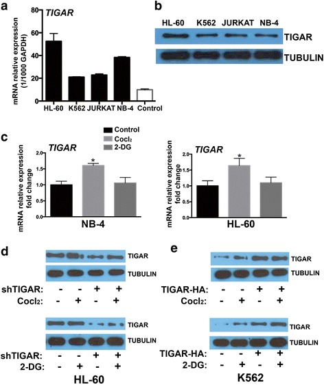
Methods: We analyzed TIGAR expression in cytogenetically normal (CN-) AML patients and the correlations with clinical and biological parameters. In vivo and in vitro, we tested whether glycolysis may induce TIGAR expression and evaluated the combination effect of glycolysis inhibitor and TIGAR knockdown on human leukemia cell proliferation.

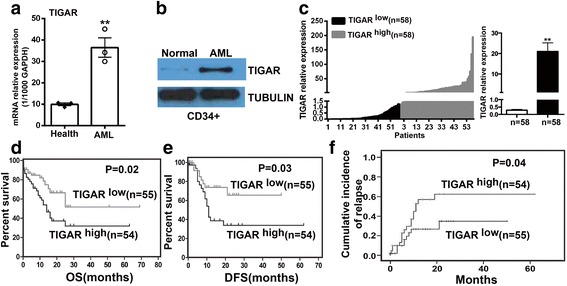
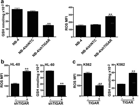
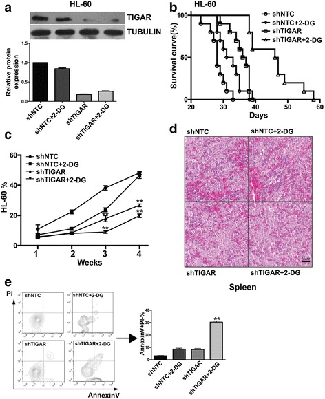
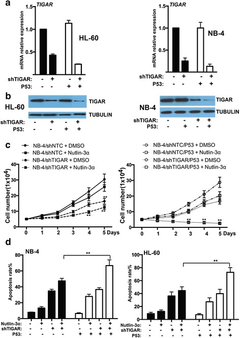
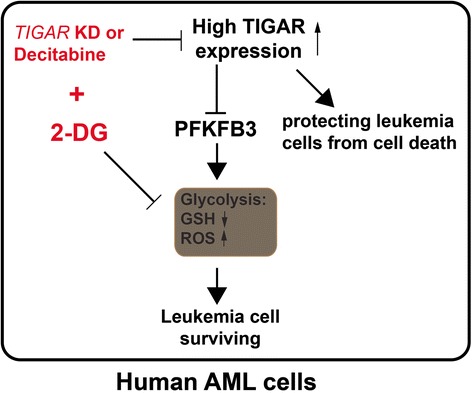
Results: High TIGAR expression was an independent predictor of poor survival and high incidence of relapse in adult patients with CN-AML. TIGAR also showed high expression in multiple human leukemia cell lines and knockdown of TIGAR activated glycolysis through PFKFB3 upregulation in human leukemia cells. Knockdown of TIGAR inhibited the proliferation of human leukemia cells and sensitized leukemia cells to glycolysis inhibitor both in vitro and in vivo. Furthermore, TIGAR knockdown in combination with glycolysis inhibitor 2-DG led leukemia cells to apoptosis. In addition, the p53 activator Nutlin-3α showed a significant combinational effect with TIGAR knockdown in leukemia cells. However, TIGAR expression and its anti-apoptotic effects were uncoupled from overexpression of exogenous p53 in leukemia cells.

Conclusions: TIGAR might be a predictor of poor survival and high incidence of relapse in AML patients, and the combination of TIGAR inhibitors with anti-glycolytic agents may be novel therapies for the future clinical use in AML patients.

Keywords: Acute myeloid leukemia; Apoptosis; Glycolysis; Survival; TIGAR.


Figures

Fig. 1
Fig. 1
TIGAR upregulation was associated with poor prognosis in AML patients. a Real-time PCR showed that TIGAR mRNA was significantly upregulated in PB cells from three AML patients in comparison with three healthy donors. The TIGAR expression was normalized to 1000 copies of GAPDH expression. b Western blotting analysis showed that the protein expression of TIGAR protein was increased in CD34+ BM cells from AML patient versus healthy donor. c Real-time PCR showed that the relative expression of TIGAR mRNA between TIGAR low and TIGAR high patients. The result represented as mean with S.E. as error bars. df Patients from TIGAR low group presented significantly longer overall survival (OS) (P = 0.021) (d) and disease-free survival (DFS) (P = 0.028) (e) and lower cumulative incidence of relapse (P = 0.044) (f) than patients from TIGAR high group
Fig. 2
Fig. 2
TIGAR showed a high expression in human leukemia cell lines, and glycolysis induced the expression of TIGAR. a, b TIGAR mRNA and protein was compared among multiple leukemia cell lines and normal cells. c TIGAR mRNA level was compared by real time PCR in both HL-60 and NB-4 cells treated with or without CoCl2 and 2-DG. d Western blotting showed the expression of TIGAR in HL-60 cells with or without TIGAR knockdown. e Western blotting showed the expression of TIGAR in K562 cells with or without TIGAR overexpression
Fig. 3
Fig. 3
TIGAR regulated the glycolysis through PFKFB3 in leukemia cells. a, b TIGAR knockdown reduced the GSH level and increased the ROS level in NB-4 cells (a) and HL-60 cells (b). c TIGAR overexpression increased the GSH level and reduced the ROS level in K562 cells
Fig. 4
Fig. 4
TIGAR knockdown inhibited the proliferation of leukemia cells and sensitized leukemia cells to glycolysis inhibition in vitro. a The cell proliferation assay showed the cell growth of HL-60 cells with TIGAR knockdown in combination with Cocl2 or 2-DG. b The cell proliferation assay showed the cell proliferation of NB-4 cells with TIGAR knockdown in combination with Cocl2 or 2-DG. c, d The cell apoptosis rate was determined by FACS in both HL-60 (c) and NB-4 cells (d) with TIGAR knockdown in combination with Cocl2 or 2-DG. HL-60 and NB-4 cells with or without TIGAR knockdown were treated with Cocl2 or 2-DG. The cells were collected on day 2 post Cocl2 or 2-DG treatment, and the apoptotic cells were determined by FACS
Fig. 5
Fig. 5
TIGAR knockdown sensitized HL-60 leukemia cells to glycolysis inhibition in vivo. a Western blotting analysis of HL-60 xenograft tumor samples. The ascites-derived tumor cells from mice were collected and lysed at the end of the study, and western blotting analyses of TIGAR and TUBULIN were performed (n = 2 for each group). b The survival of HL-60 xenograft mice with the combined treatment of TIGAR knockdown and 2-DG. 1 × 106 HL-60 cells with or without TIGAR knockdown were inoculated into BALB/c (nu/nu) nude mice (n = 10). Those mice were treated or untreated with 2-DG (2 g/kg, PO, QD) from 1-week post implantation of HL-60 cells. c In HL-60 xenograft tumor mice, the effectiveness of TIGAR knockdown in combination with 2-DG in treating HL-60 xenograft tumor mice correlated with decreased percentages of HL-60 leukemia cells in PB. FACS analysis showed the decrease of HL-60 cells in PB of HL-60 xenograft tumor mice. Mean ± SD was shown. d Photomicrographs of hematoxylin and eosin-stained spleen sections from HL-60 xenograft tumor mice with the combined treatment of TIGAR knockdown and 2-DG. e The combined treatment of TIGAR knockdown and 2-DG induced apoptosis of HL-60 leukemia cells in mice. The HL-60 cells from ascites fluid were collected and stained with PI and Annexin-V, and the percentages of PI/Annexin-V+, representing apoptotic cells, were determined by FACS (n = 5). Mean ± SD was shown
Fig. 6
Fig. 6
TIGAR expression and its anti-apoptotic effect were uncoupled from p53 in human leukemia cells. a Real-time PCR showed that the mRNA expression of TIGAR was not affected by p53 overexpression in both HL-60 and NB-4 cells. b Western blotting showed that the protein expression of TIGAR was not affected by p53 overexpression in both HL-60 and NB-4 cells. c The cell proliferation assay showed the cell growth of NB-4 cells with TIGAR knockdown in combination with p53 overexpression or/and MDM2 inhibitor Nutlin-3α. d TIGAR knockdown in combination with p53 overexpression or/and MDM2 inhibitor Nutlin-3α induced the apoptosis of NB-4 and HL-60 cells in vitro
Fig. 7
Fig. 7
A working model of TIGAR regulating the glycolysis and the proliferation of human leukemia cells. TIGAR played a dual role in human AML cells. It protected the leukemia cells from cell death while inhibited PFKFB3-regulated glycolysis in AML. Thus, TIGAR knockdown or decitabine induced apoptosis of leukemia cells through inhibiting the expression of TIGAR and sensitized leukemia cells to glycolysis inhibition in human AML

Similar articles

Cited by

References

    1. A J, Qian S, Wang G, Yan B, Zhang S, Huang Q, et al. Chronic myeloid leukemia patients sensitive and resistant to imatinib treatment show different metabolic responses. PloS one. 2010;5:e13186. - PMC - PubMed
    1. Herst PM, Howman RA, Neeson PJ, Berridge MV, Ritchie DS. The level of glycolytic metabolism in acute myeloid leukemia blasts at diagnosis is prognostic for clinical outcome. J Leukoc Biol. 2011;89:51–55. doi: 10.1189/jlb.0710417. - DOI - PubMed
    1. Zhao K, Zhou Y, Qiao C, Ni T, Li Z, Wang X, et al. Oroxylin A promotes PTEN-mediated negative regulation of MDM2 transcription via SIRT3-mediated deacetylation to stabilize p53 and inhibit glycolysis in wt-p53 cancer cells. J Hematol Oncol. 2015;8:41. doi: 10.1186/s13045-015-0137-1. - DOI - PMC - PubMed
    1. Cheung EC, Athineos D, Lee P, Ridgway RA, Lambie W, Nixon C, et al. TIGAR is required for efficient intestinal regeneration and tumorigenesis. Dev Cell. 2013;25:463–477. doi: 10.1016/j.devcel.2013.05.001. - DOI - PMC - PubMed
    1. Vander Heiden MG, Cantley LC, Thompson CB. Understanding the Warburg effect: the metabolic requirements of cell proliferation. Science. 2009;324:1029–1033. doi: 10.1126/science.1160809. - DOI - PMC - PubMed

MeSH terms