Review
. 2016 Sep;12(5):480-491.
doi: 10.1177/1745505716677139.

Neoadjuvant chemotherapy in breast cancers

Affiliations
Review

Neoadjuvant chemotherapy in breast cancers

Shahla Masood. Womens Health (Lond). 2016 Sep.

Abstract

With advances in science and technology, there are more innovations in the approach to management of patients with breast cancer. Neoadjuvant chemotherapy that is designed to be used prior to surgical removal of a tumor has received significant attention. Currently, neoadjuvant chemotherapy is offered to patients with locally advanced breast cancer and also those breast cancer patients who may benefit from size reduction before conservation therapy. There is now sufficient evidence that if neoadjuvant chemotherapy leads to complete pathologic response, the patient will enjoy a better outcome. Therefore, assessment of the degree of response to neoadjuvant chemotherapy has a major impact on patient selection and the follow-up management of each patient and defines patient outcome.

Keywords: breast cancer; breast pathology report; neoadjuvant chemotherapy.


Conflict of interest statement

Declaration of conflicting interests: The author(s) declared no potential conflicts of interest with respect to the research, authorship, and/or publication of this article.

Figures

Figure 1.
Figure 1.
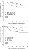
Survival rates according to stratification based on (a) IHC/FISH for ER, PR, and HER2 and (b) molecular subtyping using BluePrint and MammaPrint. Source: Reprinted with permission from Gluck et al. IHC: immunohistochemistry; FISH: fluorescence in situ hybridization; ER: estrogen receptor; PR: progesterone receptor; HER2: human epidermal growth factor receptor-2.
Figure 2.
Figure 2.
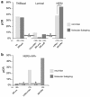
(a) pCR rates and major subtype reassignments for patients classified by BluePrint/MammaPrint molecular subtyping compared with IHC/FISH assessed subgroups. The analysis includes only patients treated with NCT (n = 403). The two major subtype reassignments were (A) conventional luminal (HR+/HER2−) patients, 35 of 188 (19%) patients reclassified by BluePrint as basal (arrow A) and (B) conventional HER2+ patients, 36 of 123 (29%) reclassified by BluePrint as luminal (arrow B). (b) pCR rates and major subtype reassignments for conventional HER2+/HR+ patients (“triple positive”) patients (95% treated with NCT/trastuzumab). A total of 36 of 75 (48%) of conventional HER2+/HR+ patients were reclassified by BluePrint as luminal—with only 1 pCR (3%) to NCT (arrow A). A total of 33 of 75 (44%) of conventional HER2+/HR+ patients were classified by BluePrint has HER2, with a pCR rate to NCT of 45% (arrow B). A total of six conventional HER2+/HR+ patients were reassigned to BPBasal (not shown). Source: Reprinted with permission from Whitworth et al.
Figure 3.
Figure 3.
Photomicrograph of tumor bed with no residual tumor characterized by stromal fibrosis and chronic inflammatory infiltrate (H&E stain ×200).
Figure 4.
Figure 4.
An example of complete pathologic response in a 52-year-old woman with palpable mass who underwent neoadjuvant chemotherapy: (a1) the tumor measured 4.1 × 3.6 × 3.2 cm3 by breast imaging, (a2) the tumor disappeared after therapy, (b1) poorly differentiated infiltrating ductal carcinoma on core needle biopsy, and (b2) there is no residual tumor seen on post-neoadjuvant chemotherapy lumpectomy sample (H&E stain ×200 and ×400).
Figure 5.
Figure 5.
(a1) An example of partial response in a 58-year-old woman with a palpable breast lesion presenting with an enhancing mass in the left breast by imaging measuring 7.9 × 5.6 × 4.7 cm3 who underwent neoadjuvant chemotherapy. (a2) Partial response is seen by a difference in the size of the lesion. (b1) This case was diagnosed as a poorly differentiated infiltrating ductal cell carcinoma (H&E stain ×400). (b2) Residual tumor cells are seen characterized by a few clusters of tumor cells in the background of fibrosis in this case (H&E stain ×200).
Figure 6.
Figure 6.
An example of no response with progressive disease in a 59-year-old woman with a 5-cm palpable mass seen on breast imaging who underwent neoadjuvant chemotherapy. (a1 and a2) Please note the progression of this tumor and (b1 and b2) in comparison of core needle biopsy findings versus lumpectomy sample diagnosed as poorly differentiated infiltrating ductal cell carcinoma (H&E stain ×200 and ×400).
Figure 7.
Figure 7.
(a) An example of complete response to neoadjuvant chemotherapy in a lymph node positive breast cancer patient with metastatic tumor seen in H&E staining of sentinel lymph node and (b) please note the disappearance of tumor cells in the same lymph node after neoadjuvant chemotherapy (H&E stain ×400).
Figure 8.
Figure 8.
(a) An example of a HER2/neu oncogene positive breast cancer patient who underwent neoadjuvant chemotherapy and (b) please note the change to HER2/neu oncogene negative status after therapy (HER2/neu gene amplification by FISH technology).
Figure 9.
Figure 9.
(a) An example of a breast cancer patient with a tumor presenting with a high proliferation rate, as evidenced by brown nuclear staining of a majority of tumor cells, who underwent neoadjuvant chemotherapy and (b) please note the decrease in the number of proliferating cells after therapy (Ki-67, immunostain 200 × 400).
Figure 10.
Figure 10.
(a) An example of a lymph node of a 64-year-old woman with an infiltrating ductal cell carcinoma who underwent neoadjuvant chemotherapy. The routine staining of the lymph shows a small area of suspicion for residual tumor. Please see circled portion (H&E stain ×200). (b) The presence of isolated and clusters of residual tumor cells are seen in the same lymph node upon immunostaining for cytokeratin. Please note the brown membrane staining of residual tumor cells (immunostained slide ×200).

References

    1. Kuerer HM, Newman LA, Smith TL, et al. Clinical course of breast cancer patients with complete pathologic primary tumor and axillary lymph node response to doxorubicin-based neoadjuvant chemotherapy. J Clin Oncol 1999; 17(2): 460–469. - PubMed
    1. Smith IE, Lipton L. Preoperative/neoadjuvant medical therapy for early breast cancer. Lancet Oncol 2001; 2: 561–570. - PubMed
    1. Kaufmann M, Von Minckwitz G, Smith R, et al. International expert panel on the use of primary (preoperative) systemic treatment of operable breast cancer: review and recommendations. J Clin Oncol 2003; 21(13): 2600–2608. - PubMed
    1. Mauri D, Pavlidis N, Ioannidis JP. Neoadjuvant versus adjuvant systemic treatment in breast cancer: a meta-analysis. J Natl Cancer Inst 2005; 97: 188–194. - PubMed
    1. Mieog J, Van der Hage JA, Van de Velde CJ. Preoperative chemotherapy for women with operable breast cancer. Cochrane Database Syst Rev 2007; 2: CD005002 - PMC - PubMed

MeSH terms