

Normative biometry of the fetal brain using magnetic resonance imaging
- PMID: 27885428
- PMCID: PMC5504265
- DOI: 10.1007/s00429-016-1342-6
Normative biometry of the fetal brain using magnetic resonance imaging
Abstract
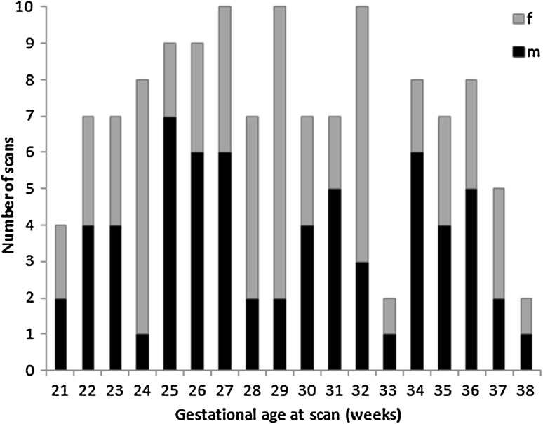
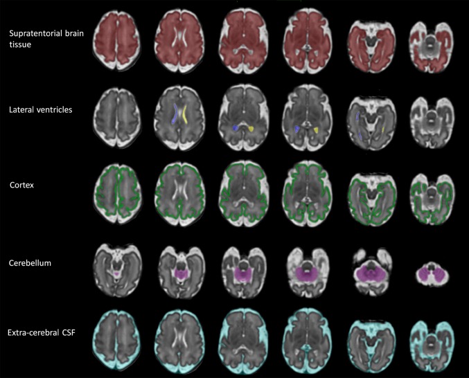
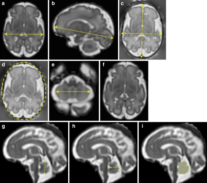
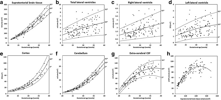
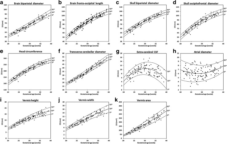
The fetal brain shows accelerated growth in the latter half of gestation, and these changes can be captured by 2D and 3D biometry measurements. The aim of this study was to quantify brain growth in normal fetuses using Magnetic Resonance Imaging (MRI) and to produce reference biometry data and a freely available centile calculator ( https://www.developingbrain.co.uk/fetalcentiles/ ). A total of 127 MRI examinations (1.5 T) of fetuses with a normal brain appearance (21-38 gestational weeks) were included in this study. 2D and 3D biometric parameters were measured from slice-to-volume reconstructed images, including 3D measurements of supratentorial brain tissue, lateral ventricles, cortex, cerebellum and extra-cerebral CSF and 2D measurements of brain biparietal diameter and fronto-occipital length, skull biparietal diameter and occipitofrontal diameter, head circumference, transverse cerebellar diameter, extra-cerebral CSF, ventricular atrial diameter, and vermis height, width, and area. Centiles were constructed for each measurement. All participants were invited for developmental follow-up. All 2D and 3D measurements, except for atrial diameter, showed a significant positive correlation with gestational age. There was a sex effect on left and total lateral ventricular volumes and the degree of ventricular asymmetry. The 5th, 50th, and 95th centiles and a centile calculator were produced. Developmental follow-up was available for 73.1% of cases [mean chronological age 27.4 (±10.2) months]. We present normative reference charts for fetal brain MRI biometry at 21-38 gestational weeks. Developing growth trajectories will aid in the better understanding of normal fetal brain growth and subsequently of deviations from typical development in high-risk pregnancies or following premature delivery.
Keywords: Brain development; Centiles; Magnetic resonance imaging.
Figures
References
-
- Almog B, Gamzu R, Achiron R, Fainaru O, Zalel Y (2003) Fetal lateral ventricular width: what should be its upper limit? A prospective cohort study and reanalysis of the current and previous data Ultrasound 39–43 - PubMed
-
- Bayley N (2006) Bayley Scales of Infant and Toddler Development. 3rd edn. Psychological Corporation - PubMed
-
- Clouchoux C, Guizard N, Evans AC, du Plessis AJ, Limperopoulos C. Normative fetal brain growth by quantitative in vivo magnetic resonance imaging AmJ. Obstet Gynecol. 2011;206:173–178. - PubMed
MeSH terms
Grants and funding
LinkOut - more resources
Full Text Sources
Other Literature Sources
Medical
