. 2017 Feb;24(2):357-370.
doi: 10.1038/cdd.2016.133. Epub 2016 Nov 25.

Induction of tumor apoptosis through a circular RNA enhancing Foxo3 activity

Affiliations

Induction of tumor apoptosis through a circular RNA enhancing Foxo3 activity

William W Du et al. Cell Death Differ. 2017 Feb.

Abstract

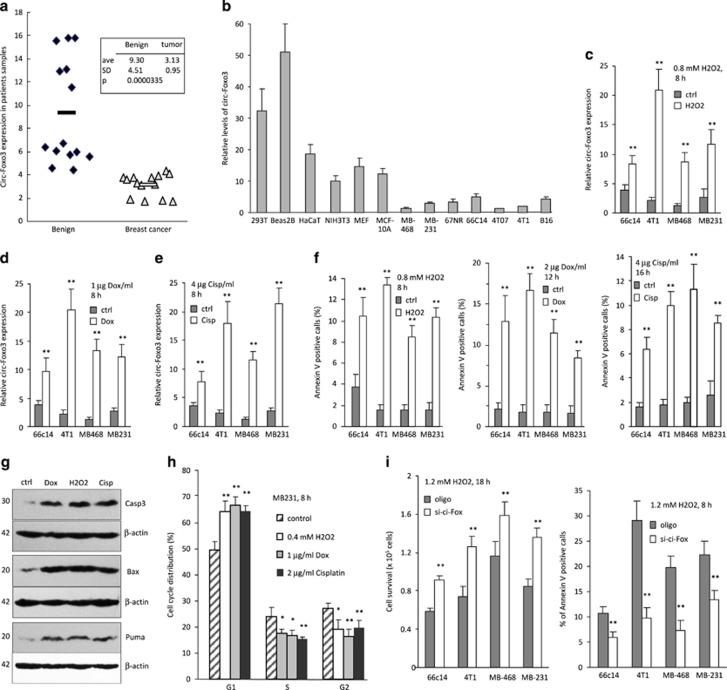
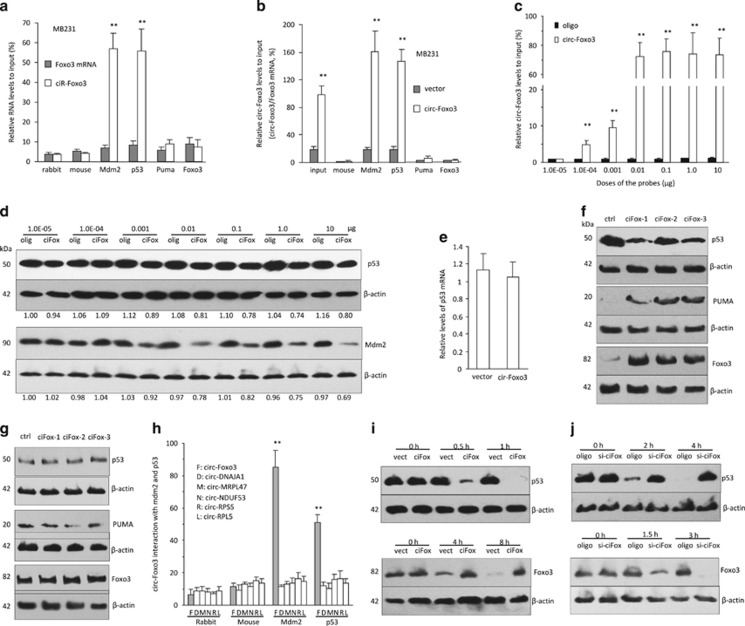
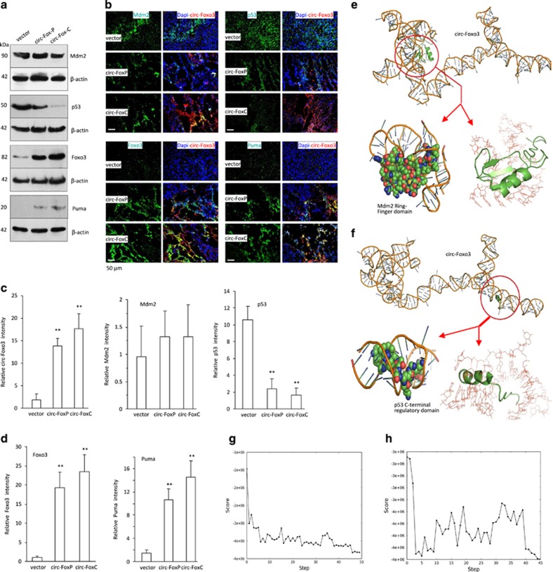
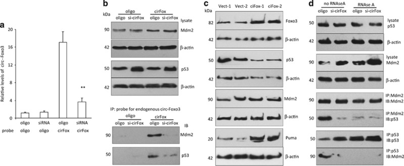
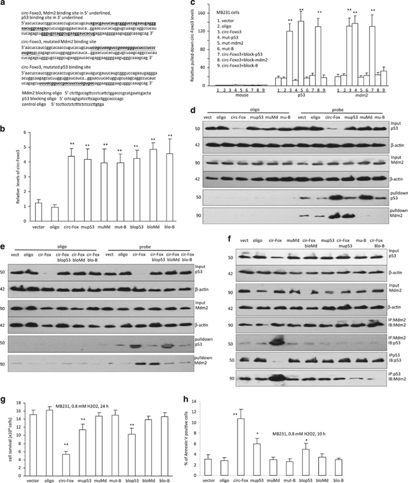
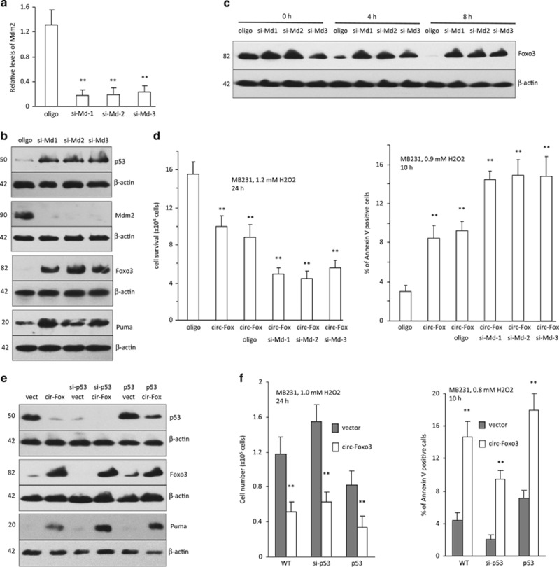
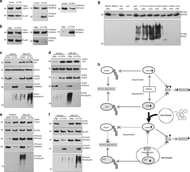
Circular RNAs are a class of non-coding RNAs that are receiving extensive attention. Despite reports showing circular RNAs acting as microRNA sponges, the biological functions of circular RNAs remain largely unknown. We show that in patient tumor samples and in a panel of cancer cells, circ-Foxo3 was minimally expressed. Interestingly, during cancer cell apoptosis, the expression of circ-Foxo3 was found to be significantly increased. We found that silencing endogenous circ-Foxo3 enhanced cell viability, whereas ectopic expression of circ-Foxo3 triggered stress-induced apoptosis and inhibited the growth of tumor xenografts. Also, expression of circ-Foxo3 increased Foxo3 protein levels but repressed p53 levels. By binding to both, circ-Foxo3 promoted MDM2-induced p53 ubiquitination and subsequent degradation, resulting in an overall decrease of p53. With low binding affinity to Foxo3 protein, circ-Foxo3 prevented MDM2 from inducing Foxo3 ubiquitination and degradation, resulting in increased levels of Foxo3 protein. As a result, cell apoptosis was induced by upregulation of the Foxo3 downstream target PUMA.


Conflict of interest statement

The authors declare no conflict of interest.

Figures

Figure 1
Figure 1
The effect of circ-Foxo3 on cell apoptosis. (a) Total RNAs were isolated from the specimens of patients with breast carcinoma and subject to real-time PCR measurement. Tumor samples showed significantly lower levels of circ-Foxo3 than the benign samples. (b) Expression of circ-Foxo3 was analyzed in a variety of cell lines by real-time PCR. Six non-cancer cell lines expressed significantly higher levels of circ-Foxo3 than seven cancer cell lines. (ce) Cancer cell lines 66C14, 4T1, MDA-MB-468, and MDA-MB-231 were cultured in the presence of H2O2 (c), Dox (d), or Cisplatin (e). RNAs were isolated and subject to real-time PCR to measure circ-Foxo3 levels. Asterisks indicate significant differences.*P<0.05, **P<0.01. Error bars, S.D. (n=3). (f) The cells were subject to Annexin-V staining to detect apoptosis. Treatment with H2O2 (left), Dox (middle), or Cisplatin (right) increased apoptosis. (g) Western blot showed that H2O2, Dox and Cisp treatment enhanced Bax and Puma expression and increased levels of cleaved Caspase-3 in MB-231 cells. (h) Cell cycle analysis showed that H2O2, Dox and Cisp treatment repressed cell cycle entry. *P<0.05, **P<0.01. Error bars, S.D. (n=4). (i) 66C14, 4T1, MB-468, and MB-231 cells were transfected with circ-Foxo3 siRNA or a control oligo. The cells were cultured in basal medium with 1.2 mM H2O2 for 18 h for survival assay (left) or maintained in 1.2 mM H2O2 for 8 h and subject to Annexin-V staining for apoptosis assay (right). Transfection with the circ-Foxo3 siRNA increased cell survival (left) and decreased apoptosis (right). *P<0.05, **P<0.01. Error bars, S.D. (n=3)
Figure 2
Figure 2
Expression of circ-Foxo3 inhibited tumor growth and enhanced mouse survival. (a) Cultured in serum-free medium for 6 days, the circ-Foxo3-transfected 4T1 cells showed decreased cell survival compared with mock control cells. *P<0.05, **P<0.01. Error bars, S.D. (n=6). (b) The transfected 4T1 cells were cultured in basal medium with H2O2 for 16 h for survival assay. *P<0.05, **P<0.01. Error bars, S.D. (n=6). (c) The transfected 4T1 cells were cultured in serum-free medium for 48 h, and subjected to Annexin-V staining, followed by flow cytometry to measure apoptotic cells. *P<0.05, **P<0.01. Error bars, S.D. (n=4). (d) The cells were cultured in basal medium with 0.6 mM H2O2 for 6 h, and subjected to Annexin-V staining, followed by flow cytometry to measure apoptotic cells. *P<0.05, **P<0.01. Error bars, S.D. (n=4). (e) The circ-Foxo3- and mock-transfected cells 66C14, 4T1, MDA-MB-468, and MDA-MB-231 were cultured in serum-free medium for 6 days for cell survival assay. The circ-Foxo3-transfected cell lines showed decreased viability compared with the control. *P<0.05, **P<0.01. Error bars, S.D. (n=3). (f) The cells were cultured in serum-free medium for 3 days, followed by apoptotic assay. circ-Foxo3 expression enhanced apoptosis. *P<0.05, **P<0.01. Error bars, S.D. (n=3). (gh) 4T1 cells transfected with circ-Foxo3 and the vector were subject to tumorigenesis assay. Expression of circ-Foxo3 inhibited tumor growth (g). Typical photo of tumors (h). (i) Tumor sections were subject to TUNEL staining followed by quantification of the TUNEL-positive cells. Significantly higher levels of cell death were detected in the tumors formed by cells transfected with circ-Foxo3 compared with the control. (j) Western blot showed that the tumor tissues expressing high levels of circ-Foxo3 had increased levels of Bax, Puma, and cleaved Caspase-3. (k) Mice were intraperitoneally injected with B16 cells with a control vector or circ-Foxo3 plasmid, and with circ-Foxo3 transfected B16 cells, followed by survival test. Kaplan–Meier curve was obtained. Injection of circ-Foxo3 plasmid or circ-Foxo3-transfected B16 cells increased mouse survival significantly (**P<0.01). (l) PCR showed that circ-Fox3 expressed at high levels in plasmid injection and circ-Foxo3-transfected groups of tumors. (m) In TUNEL staining, significantly higher levels of cell death were detected in the tumors of mice injected with circ-Foxo3 plasmids or circ-Foxo3-transfected cells relative to the control. (n) PCR showed that normal mouse breast tissues expressed high levels of circ-Foxo3 compared with 4T1 tumors and cell lines. *P<0.05, **P<0.01. Error bars, S.D. (n=4)
Figure 3
Figure 3
Expression of circ-Foxo3 decreased p53 but increased Foxo3 levels. (a) MB-231 cell lysis were subject to immunoprecipitation with anti-rabbit IgG, mouse IgG, MDM2, p53, Puma, and Foxo3 antibodies, followed by real-time PCR with primers specific for Foxo3 or circ-Foxo3. Antibodies against MDM2 and p53 pulled down circ-Foxo3, but not Foxo3 mRNA. **P<0.01. Error bars, S.D. (n=4). (b) Cell lysates prepared from MB-231 cells transfected with circ-Foxo3 or the mock control were subject to immunoprecipitation with different antibodies, followed by real-time PCR. Antibodies against MDM2 and p53 pulled down more circ-Foxo3 from the circ-Foxo3-transfected cells than from the control cells. **P<0.01. Error bars, S.D. (n=4). (c) Lysates prepared from BEAS2B cells were hybridized with biotinylated probe with increased concentration, and subject to RNA pull-down assays. Real-time PCR showed that circ-Foxo3 pulled down high levels of circ-Foxo3, reaching plateau at the dose of 10−2μg. **P<0.01. Error bars, S.D. (n=4). (d) After being pulled down by the circ-Foxo3 probe, the remaining lysates were subject to western blotting. Both MDM2 and p53 showed decreased levels after circ-Foxo3 was pulled down with the circ-Foxo3 probe at the doses over 10−2μg. (e) PCR showed that overexpression of circ-Foxo3 did not change p53 mRNA levels. (f) Lysates prepared from MB-231 cells transfected with mock control or circ-Foxo3, were subject to western blot analysis. Expression of circ-Foxo3 decreased p53 levels, but increased Puma and Foxo3 levels. (g) The circ-Foxo3- and vector-transfected MB-231 cells were treated with 10 μM MG-132 for 12 h. Lysates were subject to western blot with antibodies against p53, Puma, and Foxo3. Little change was detected. (h) The pulled down pellets were also measured for the levels of other circular RNAs including circ-DNSJA1, circ-MRPL47, circ-NDUF53, circ-RPS5, and circ-PRL5. Antibodies against MDM2 and p53 could only pull-down circ-Foxo3. (i) The circ-Foxo3- and vector-transfected MB-231 cells were cultured in basal medium containing 20 μM Emetine for the time points as indicated, followed by western blotting. circ-Foxo3 expression decreased p53 (upper) but increased Foxo3 (lower) levels compared with mock control. (j) Lysates prepared from MB-231 cells transfected with circ-Foxo3 siRNA or a control oligo were treated with 20 μM Emetine for the time points as indicated, followed by western blotting. Transfection with circ-Foxo3 siRNA increased p53 (upper) but decreased (Foxo3) levels (lower)
Figure 4
Figure 4
Interaction of circ-Foxo3 with MDM2 and p53 (a) B16 tumor lysates were subject to western blot with antibodies to p53, Foxo3, Puma, Mdm2 and β-actin. circ-Foxo3 enhanced Foxo3 and Puma expression and repressed p53 expression in the tumors. (b) Tumor sections were stained with DAPI (blue), circ-Foxo3 (red, by in situ hybridization) and related proteins including Mdm2, p53, Foxo3, and Puma (green, by immunohistochemical staining). Typical photos showed that expression of circ-Foxo3 was colocalized with these protein. (c) Quantification of circ-Foxo3, Mdm2, and p53 in the tumor sections. (d) Quantification of Foxo3 and Puma in the tumor sections. (e) Graphical representation of three-dimensional structures of circ-Foxo3 and MDM2 (RING-finger domain) docking models with a zoom-in image of the binding interface done by NPDock. The binding region is shown in two different visualizations (cartoon and sphere). (f) Graphical representation of three-dimensional structures of circ-Foxo3 and p53 (C-terminal regulatory domain) docking models with a zoom-in image of the binding interface done by NPDock. The binding region is shown in two different visualizations (cartoon and sphere). (g) Refinement of the best docked circ-Foxo3-MDM2 (RING-finger domain) model showing MC score vs steps of simulation. (h) Refinement of the best docked circ-Foxo3-p53 (C-terminal regulatory domain) model showing MC score vs steps of simulation. *P<0.05, **P<0.01
Figure 5
Figure 5
The interaction of circ-Foxo3 with p53 and MDM2. (a) Lysates prepared from MB-231 cells transfected with circ-Foxo3 siRNA or a control oligo were hybridized with the biotinylated probe. Real-time PCR showed that circ-Foxo3 probe pulled down less circ-Foxo3 in the circ-Foxo3 siRNA-transfected cells. **P<0.01. Error bars, S.D. (n=4). (b) Upper, the lysates were subject to western blot with antibodies against MDM2 and p53. Increased p53 levels were detected. Lower, the lysates were subject to RNA pull-down assays. The circ-Foxo3 probe pulled down less MDM2 and p53 in the cells transfected with the circ-Foxo3 siRNA. (c) The tumors formed by the circ-Foxo3-transfected cells revealed higher levels of Foxo3 and Puma, but lower levels of p53 compared with the controls. There was little difference in the levels of MDM2. (d) Lysates prepared from MB-231 cells transfected with circ-Foxo3 siRNA or a control oligo were incubated with or without 1 μg/ml RNAse-A for 30 min. Antibody against MDM2 pulled down decreased levels of p53 and antibody against p53 pulled down decreased levels of MDM2, when circ-Foxo3 was silenced or treated with RNAse-A
Figure 6
Figure 6
Mutation of circ-Foxo3 in the MDM2 and p53-binding sites. (a) Sequences of circ-Foxo3 with Mdm2 and p53-binding sites, designed mutated binding sites, and blocking oligos. (b) PCR showed the expression of circ-Foxo3 in cell lysates prepared from MB-231 cells transfected with the control vector, oligo, circ-Foxo3, circ-Foxo3 mutated in p53-binding sites (mut-p53), circ-Foxo3 mutated in Mdm2-binding sites (mut-mdm2), circ-Foxo3 mutated in both p53 and Mdm2-binding sites (mut-B), circ-Foxo3 and p53 blocking oligo (block-p53), circ-Foxo3 and Mdm2 blocking oligo (block-mdm2), and circ-Foxo3 and both p53 and Mdm2 blocking oligos (block-B). (c) Real-time PCR showed that antibodies against MDM2 and p53 pulled down more circ-Foxo3 from the circ-Foxo3-transfected cells than from the control cells, but not in the binding site mutated cells or cells treated with blocking oligos. **P<0.01. Error bars, S.D. (n=4). (d) Cell lysates prepared from MB-231 cells transfected with the control vector (vect), oligo, circ-Foxo3, mutated p53 (mup53), mutated Mdm2 (muMd), mutated in both p53 and Mdm2-binding sites (mu-B) were subject to circ-Foxo3 pull-down assays. The circ-Foxo3 probe pulled down MDM2 and p53 of circ-Foxo3 expressing cells, but not the binding site mutated cell lines. (e) Cell lysates prepared from MB-231 cells transfected with the vector, oligo, circ-Foxo3, circ-Foxo3 and blop53, circ-Foxo3 and bloMdm2, and circ-Foxo3 and block-B. The circ-Foxo3 probe pulled down MDM2 and p53 in circ-Foxo3 expressing cells, which was blocked by the blocking oligos. (f) Western blot showed p53 and Mdm2 expression in MB-231 cells transfected with different constructs. Precipitation of Mdm2 pulled down p53 and precipitation of p53 pulled down Mdm2, in the circ-Foxo3 overexpressing cells, but not in the binding site mutated cells nor cells co-transfected with the blocking oligos. (g) The above cells were cultured in 0.8 mM H2O2 for 24 h. Expression of circ-Foxo3 repressed cell survival, which could be prevented by binding site mutation or application of the blocking oligos. (h) The cells were cultured in 0.8 mM H2O2 for 10 h. Expression of circ-Foxo3 enhanced cell apoptosis, which could be prevented by binding site mutation or application of the blocking oligos. *P<0.05, **P<0.01. Error bars, S.D. (n=3)
Figure 7
Figure 7
circ-Foxo3 function by modulating expression of p53 and Foxo3. (a) Real-time PCR showed that three siRNAs-targeting Mdm2 decreased Mdm2 levels in MB-231 cells transfected with these siRNAs. **P<0.01. Error bars, S.D. (n=3). (b) Western blot showed that silencing Mdm2 enhanced expression of p53, Foxo3 and Puma. (c) MB-231 cells transfected with Mdm2 siRNA or a control oligo were treated with 50 μM Emetine for the time points as indicated. Silencing Mdm2 inhibited Foxo3 degradation. (d) Left, silencing MDM2 decreased cell survival. **P<0.01. Error bars, S.D. (n=4). Right, silencing Mdm2 increased apoptosis which showed enhanced annexin-V staining **P<0.01. Error bars, S.D. (n=4). (e) Western blot showed that transfection with circ-Foxo3 enhanced Foxo3 and Puma expression with p53 silencing or p53 overexpression. (f) Left, transfection with circ-Foxo3 repressed cell survival with p53 silencing or p53 overexpression. **P<0.01. Error bars, S.D. (n=4). Right, transfection with circ-Foxo3 increased cell apoptosis with p53 silencing or p53 overexpression. The cells were cultured in 0.9 mM H2O2 for 10 h, showing that expression of circ-Foxo3 **P<0.01. Error bars, S.D. (n=4)
Figure 8
Figure 8
Expression of circ-Foxo3 modulated the function of MDM2 in ubiquitination of p53 and Foxo3. (a) Circ-Foxo3 expression had no effect on MDM2 levels. MDM2 precipitation pulled down more p53 and less Foxo3 in the circ-Foxo3-transfected cells than in the mock control. (b) Transfection with circ-Foxo3 siRNA did not affect MDM2 expression. MDM2 precipitation pulled down more Foxo3 but less p53 in the circ-Foxo3 siRNA-transfected cells. (c) circ-Foxo3 transfection decreased p53 level. Treatment with 10 μM MG-132 for 12 h abolished this effect. p53 precipitation pulled down more MDM2 and ubiquitin (detected with antibody against ubiquintin from Cell signalling, Cat No. 3933) in the circ-Foxo3-transfected cells. (d) Transfection with circ-Foxo3 siRNA increased p53 levels, which was abolished by treatment with MG-132. p53 precipitation pulled down less MDM2 and ubiquitin in the circ-Foxo3 siRNA-transfected cells. (e) Foxo3 precipitation pulled down less MDM2 and ubiquitin in the circ-Foxo3 transfected cells. (f) Foxo3 precipitation pulled down more MDM2 and ubiquitin in the circ-Foxo3 siRNA-transfected cells. (g) Cell lysates prepared from MB-231 cells co-transfected with ubiquintin and mock control, circ-Foxo3, mup53, muMd or mu-B, were subject to precipitation with antibody against ubiquintin. Precipitation of ubiquintin pulled down p53 in the circ-Foxo3-overexpressing cell lines. (h) MDM2 acts as an E3 ubiquitin ligase, targeting both p53 and Foxo3 for degradation in proteasome. MDM2 is capable of poly-ubiquitinating p53 and Foxo3. circ-Foxo3 interacts with both MDM2 and p53, which enhances the interaction between MDM2 and p53, and further increases the poly-ubiquitination and degradation of p53. Another target protein of MDM2, Foxo3, is free from being degraded. Thus, overexpression of circ-Foxo3, decreases the interaction between Foxo3 and MDM2, decreasing the amount of poly-ubiquitination and degradation of Foxo3. Increased Foxo3 then promotes expression of Puma, and enhances cell apoptosis

Comment in

References

    1. Jeck WR, Sharpless NE. Detecting and characterizing circular RNAs. Nat Biotechnol 2014; 32: 453–461. - PMC - PubMed
    1. Haimovich G, Medina DA, Causse SZ, Garber M, Millan-Zambrano G, Barkai O et al. Gene expression is circular: factors for mRNA degradation also foster mRNA synthesis. Cell 2013; 153: 1000–1011. - PubMed
    1. Wilusz JE, Sharp PA. Molecular biology. A circuitous route to noncoding RNA. Science 2013; 340: 440–441. - PMC - PubMed
    1. Westholm JO, Miura P, Olson S, Shenker S, Joseph B, Sanfilippo P et al. Genome-wide analysis of drosophila circular RNAs reveals their structural and sequence properties and age-dependent neural accumulation. Cell Rep 2014; 9: 1966–1980. - PMC - PubMed
    1. Zhang XO, Wang HB, Zhang Y, Lu X, Chen LL, Yang L. Complementary sequence-mediated exon circularization. Cell 2014; 159: 134–147. - PubMed

MeSH terms

Grants and funding