

Expression of Barhl2 and its relationship with Pax6 expression in the forebrain of the mouse embryo
- PMID: 27887593
- PMCID: PMC5124293
- DOI: 10.1186/s12868-016-0311-6
Expression of Barhl2 and its relationship with Pax6 expression in the forebrain of the mouse embryo
Abstract
Background: The transcription factor Barhl2 is an antiproneural transcription factor with roles in neuronal differentiation. The functions of its homologue in Drosophila development are better understood than its functions in mammalian brain development. Existing evidence suggests that its expression in the embryonic forebrain of the mouse is regional and may complement that of another transcription factor that is important for forebrain development, Pax6. The aim of this study is to provide a more detailed description of the Barhl2 expression pattern in the embryonic forebrain than is currently available, to relate its expression domains to those of Pax6 and to examine the effects of Pax6 loss on Barhl2 expression.
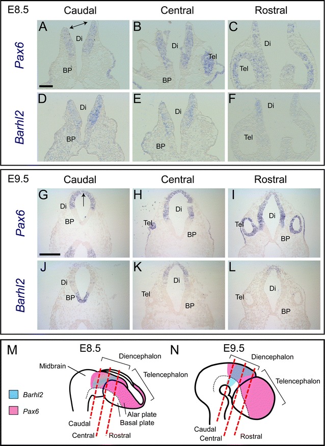
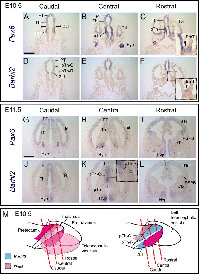
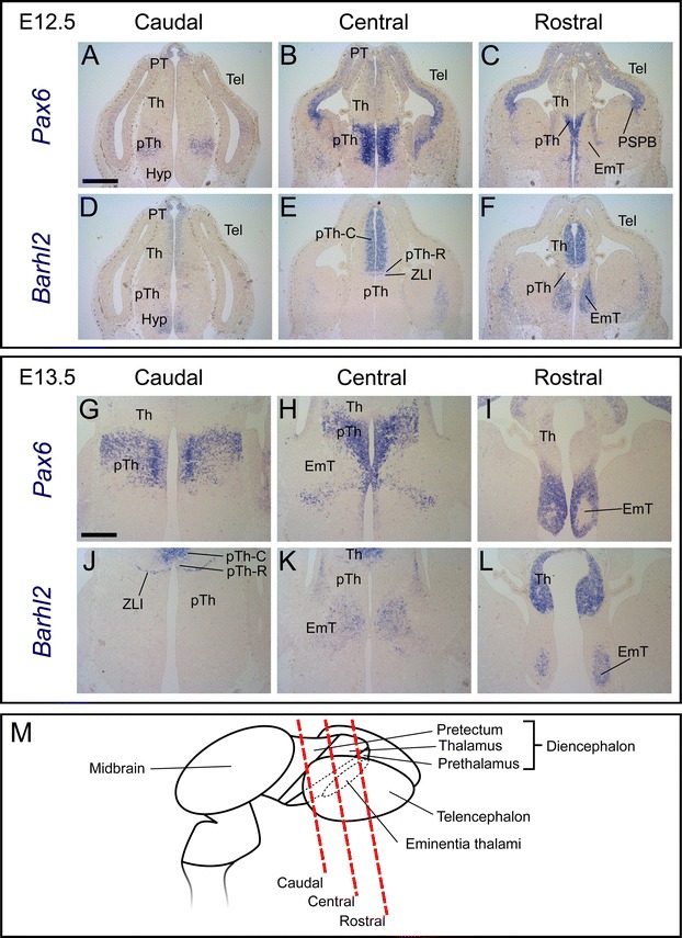
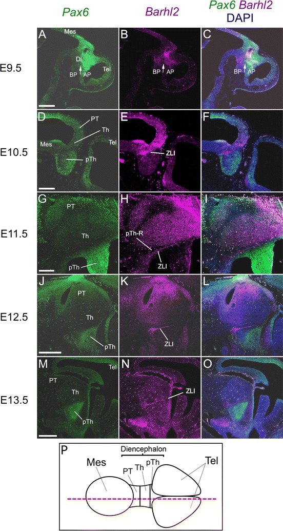
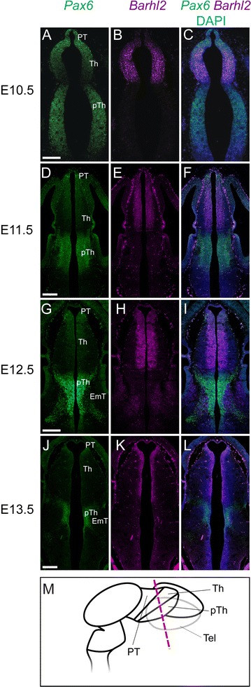
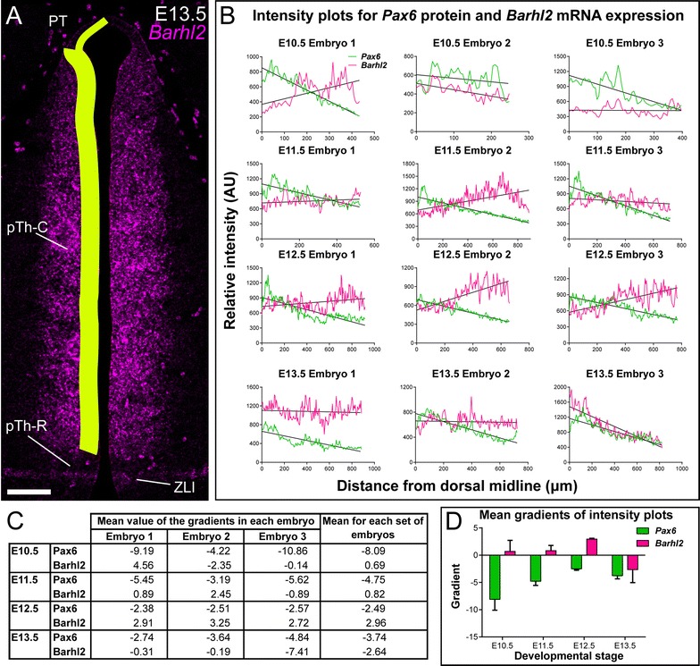
Results: We found that Barhl2 is expressed in the developing diencephalon from the time of anterior neural tube closure. Its expression initially overlaps that of Pax6 in a central region of the alar diencephalon but over the following days their domains of expression become complementary in most forebrain regions. The exceptions are the thalamus and pretectum, where countergradients of Pax6 and Barhl2 expression are established by embryonic day 12.5, before overall Pax6 levels in these regions decline greatly while Barhl2 levels remain relatively high. We found that Barhl2 expression becomes upregulated in specifically the thalamus and pretectum in Pax6-null mice.
Conclusions: The region-specific expression pattern of Barhl2 makes it likely to be an important player in the development of region-specific differences in embryonic mouse forebrain. Repression of its expression in the thalamus and pretectum by Pax6 may be crucial for allowing proneural factors to promote normal neuronal differentiation in this region.
Keywords: Barhl2; Development; Forebrain; Gene expression; Mouse; Pax6; Thalamus; Zona limitans intrathalamica.
Figures


Similar articles
-
Emx2 and Pax6 function in cooperation with Otx2 and Otx1 to develop caudal forebrain primordium that includes future archipallium.J Neurosci. 2005 May 25;25(21):5097-108. doi: 10.1523/JNEUROSCI.0239-05.2005. J Neurosci. 2005. PMID: 15917450 Free PMC article.
-
Barhl2 Determines the Early Patterning of the Diencephalon by Regulating Shh.Mol Neurobiol. 2017 Aug;54(6):4414-4420. doi: 10.1007/s12035-016-0001-5. Epub 2016 Jun 27. Mol Neurobiol. 2017. PMID: 27349434 Free PMC article.
-
The conserved barH-like homeobox-2 gene barhl2 acts downstream of orthodentricle-2 and together with iroquois-3 in establishment of the caudal forebrain signaling center induced by Sonic Hedgehog.Dev Biol. 2014 Dec 1;396(1):107-20. doi: 10.1016/j.ydbio.2014.09.027. Epub 2014 Oct 2. Dev Biol. 2014. PMID: 25281935
-
Role of Pax6 in forebrain regionalization.Brain Res Bull. 2005 Sep 15;66(4-6):387-93. doi: 10.1016/j.brainresbull.2005.02.006. Epub 2005 Feb 24. Brain Res Bull. 2005. PMID: 16144620 Review.
-
The role of Pax6 in forebrain development.Dev Neurobiol. 2011 Aug;71(8):690-709. doi: 10.1002/dneu.20895. Dev Neurobiol. 2011. PMID: 21538923 Review.
Cited by
-
The role of the diencephalon in the guidance of thalamocortical axons in mice.Development. 2020 Jun 26;147(12):dev184523. doi: 10.1242/dev.184523. Development. 2020. PMID: 32541009 Free PMC article.
-
Small RNA-Seq reveals novel miRNAs shaping the transcriptomic identity of rat brain structures.Life Sci Alliance. 2018 Sep 30;1(5):e201800018. doi: 10.26508/lsa.201800018. eCollection 2018 Oct. Life Sci Alliance. 2018. PMID: 30456375 Free PMC article.
-
Transcriptome analysis reveals determinant stages controlling human embryonic stem cell commitment to neuronal cells.J Biol Chem. 2017 Dec 1;292(48):19590-19604. doi: 10.1074/jbc.M117.796383. Epub 2017 Sep 26. J Biol Chem. 2017. PMID: 28972157 Free PMC article.
-
Development of the early fetal human thalamus: from a protomap to emergent thalamic nuclei.Front Neuroanat. 2025 Feb 7;19:1530236. doi: 10.3389/fnana.2025.1530236. eCollection 2025. Front Neuroanat. 2025. PMID: 39990522 Free PMC article.
-
Spatiotemporal transcriptomic maps of whole mouse embryos at the onset of organogenesis.Nat Genet. 2023 Jul;55(7):1176-1185. doi: 10.1038/s41588-023-01435-6. Epub 2023 Jul 6. Nat Genet. 2023. PMID: 37414952 Free PMC article.
References
Publication types
MeSH terms
Substances
Grants and funding
LinkOut - more resources
Full Text Sources
Other Literature Sources
Molecular Biology Databases
