Immunohistochemical insights into Saffold virus infection of the brain of juvenile AG129 mice
- PMID: 27887630
- PMCID: PMC5123230
- DOI: 10.1186/s12985-016-0654-8
Immunohistochemical insights into Saffold virus infection of the brain of juvenile AG129 mice
Abstract
Background: Saffold Virus (SAFV) is a human cardiovirus that is suspected of causing infection of the central nervous system (CNS) in children. While recent animal studies have started to elucidate the pathogenesis of SAFV, very little is known about the mechanisms behind it.
Method: In this study, we attempted to elucidate some of the mechanisms of the pathogenesis of SAFV in the brain of a juvenile mouse model by using immunohistochemical methods.
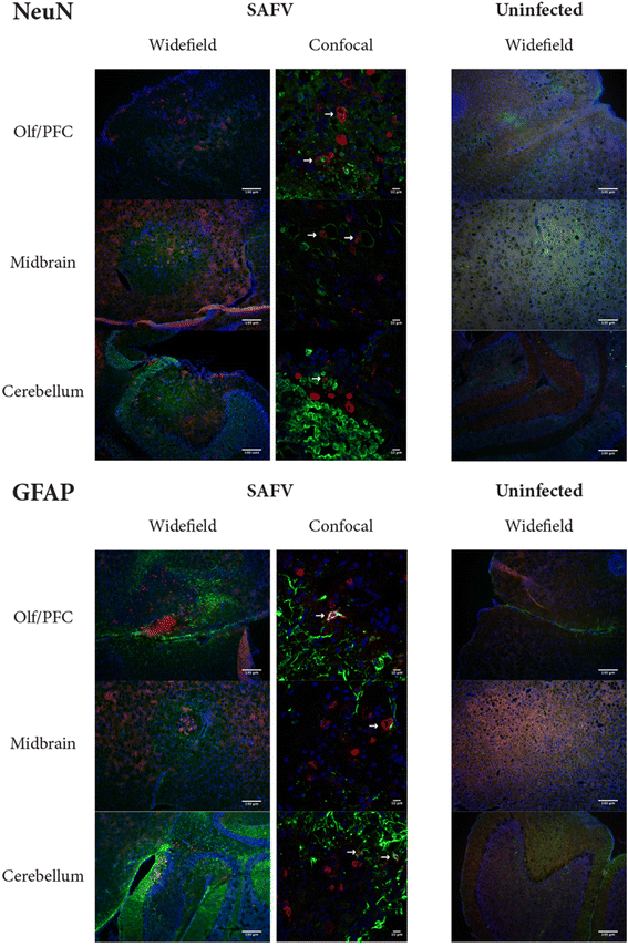
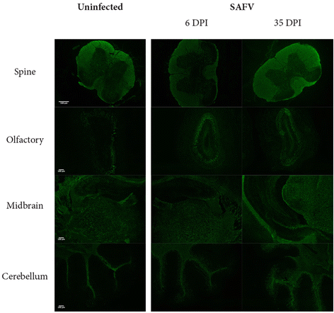
Results: We first showed that SAFV is able to infect both neuronal and glial cells in the brain of 2 week-old AG129 mice. We then showed that SAFV is able to induce apoptosis in both neuronal and glial cells in the brain. Lastly, we showed that SAFV infection does not show any signs of gross demyelination in the brain.
Conclusion: Overall, our results provide important insights into the mechanisms of SAFV in the brain.
Keywords: Apoptosis; Demyelination; Juvenile brain; Saffold virus.
Figures


Similar articles
-
The Pathogenesis of Saffold Virus in AG129 Mice and the Effects of Its Truncated L Protein in the Central Nervous System.Viruses. 2016 Feb 18;8(2):24. doi: 10.3390/v8020024. Viruses. 2016. PMID: 26901216 Free PMC article.
-
Neurotropism of Saffold virus in a mouse model.J Gen Virol. 2016 Jun;97(6):1350-1355. doi: 10.1099/jgv.0.000452. Epub 2016 Mar 9. J Gen Virol. 2016. PMID: 26959376
-
Saffold virus, an emerging human cardiovirus.Rev Med Virol. 2017 Jan;27(1):e1908. doi: 10.1002/rmv.1908. Epub 2016 Oct 10. Rev Med Virol. 2017. PMID: 27723176 Free PMC article. Review.
-
Neuropathogenicity of Two Saffold Virus Type 3 Isolates in Mouse Models.PLoS One. 2016 Feb 1;11(2):e0148184. doi: 10.1371/journal.pone.0148184. eCollection 2016. PLoS One. 2016. PMID: 26828718 Free PMC article.
-
Saffold virus, a novel human Cardiovirus with unknown pathogenicity.J Virol. 2012 Feb;86(3):1292-6. doi: 10.1128/JVI.06087-11. Epub 2011 Nov 23. J Virol. 2012. PMID: 22114344 Free PMC article. Review.
References
Publication types
MeSH terms
LinkOut - more resources
Full Text Sources
Other Literature Sources

