. 2017 Jan 15;130(2):332-343.
doi: 10.1242/jcs.195305. Epub 2016 Nov 25.

Drosophila Sulf1 is required for the termination of intestinal stem cell division during regeneration

Affiliations

Drosophila Sulf1 is required for the termination of intestinal stem cell division during regeneration

Masahiko Takemura et al. J Cell Sci. .

Abstract

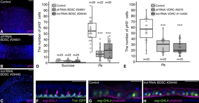
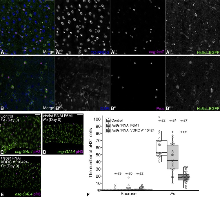
Stem cell division is activated to trigger regeneration in response to tissue damage. The molecular mechanisms by which this stem cell mitotic activity is properly repressed at the end of regeneration are poorly understood. Here, we show that a specific modification of heparan sulfate is crucial for regulating Drosophila intestinal stem cell (ISC) division during normal midgut homeostasis and regeneration. Loss of the extracellular heparan sulfate endosulfatase Sulf1 resulted in increased ISC division during normal homeostasis, which was caused by upregulation of mitogenic signaling including the JAK-STAT, EGFR and Hedgehog pathways. Using a regeneration model, we found that ISCs failed to properly halt division at the termination stage in Sulf1 mutants, showing that Sulf1 is required for terminating ISC division at the end of regeneration. We propose that post-transcriptional regulation of mitogen signaling by heparan sulfate structural modifications provides a new regulatory step for precise temporal control of stem cell activity during regeneration.

Keywords: Drosophila; Heparan sulfate proteoglycan; Intestine; Regeneration.


Conflict of interest statement

The authors declare no competing or financial interests.

Figures

Fig. 1.
Fig. 1.
Heparan sulfate is required for ISC division during regeneration. (A–C) Flies with esgts driving UAS-sfl RNAi BDSC #34601 (B) or UAS-trol RNAi BDSC #29440 (C) were infected with P. entomophila (Pe) followed by immunostaining for pH3 (magenta). esgts midgut without a UAS transgene was used as a control (A). (D,E) Quantification of pH3+ cells in the posterior midgut of indicated genotypes after mock or P. entomophila infection. The boxes represent the interquartile range, and the line represents the median. The whiskers extend to the highest and lowest values within 1.5 times the interquartile range. One outlier is not shown in the graph for better visualization but is included in the statistical analysis (D). (F) Optical cross section of a posterior midgut showing progenitor cells (ISCs and enteroblasts, marked with esg-GAL4/UAS-tdTomato, magenta). F-actin is labeled with phalloidin (blue). Trol::GFP (green) is deposited in the basement membrane. (G,H) Optical cross sections of the control midgut (G) and the midgut depleted of trol in progenitor cells (H). Phalloidin staining is shown in magenta. TO-PRO-3 was used to stain nuclei (blue). ***P<0.001 (Wilcoxon rank-sum test). Scale bars: 50 µm (A–C), 25 µm (F–H).
Fig. 2.
Fig. 2.
Heparan sulfate 6-O sulfation is required for ISC division during regeneration. (A) A posterior midgut from a 7–8-day-old female fly carrying esg-lacZ (ISCs and enteroblasts, magenta) and Hs6st::EGFP (green). TO-PRO-3 was used to stain nuclei (blue). (B) A posterior midgut showing Hs6st::EGFP expression (green), Pros (magenta), and nuclei (DAPI, blue). (C–E) Posterior midgut samples of esgts (green) with UAS-Hs6st RNAi F6M1 (D) or UAS-Hs6st RNAi VDRC #110424 (E) were infected with P. entomophila (Pe) followed by immunostaining for pH3 (magenta). esgts midgut without a RNAi transgene was used as a control (C). (F) Quantification of pH3+ cells per posterior midgut. The boxes represent the interquartile range, and the line represents the median. The whiskers extend to the highest and lowest values within 1.5 times the interquartile range. *P<0.05; ***P<0.001 (Wilcoxon rank-sum test). Scale bars: 25 µm (A,B); 50 µm (C–E).
Fig. 3.
Fig. 3.
Generation of a novel Sulf1 null allele using the CRISPR/Cas9 system. (A) Two single-guide RNAs (sgRNAs) were designed to induce double-strand breaks (DSBs) near the Sulf1 translational start (ATG in green) and stop sites (TAA in green), respectively. The sgRNA targets and the protospacer adjacent motifs (PAMs) are shown in red and cyan, respectively. (B) The sgRNAs were injected into fly embryos expressing Cas9 in the germline under the control of the vasa regulatory sequences (Gratz et al., 2014), along with a repair template that contains a coding sequence for mCherry, flanked by 1.2-kb Sulf1 homology arms. In the Sulf1{KO; mCherry} allele, most of the Sulf1 coding sequence (black) is replaced with the mCherry coding sequence (magenta). (C) Sanger sequencing reads of Sulf1{KO; mCherry} genomic DNA. (D) A wing disc homozygous for Sulf1{KO; mCherry} was immunostained for mCherry (magenta). TO-PRO-3 was used for nuclear counterstaining (blue). Scale bar: 50 µm (D).
Fig. 4.
Fig. 4.
Sulf1 is expressed in enterocytes and visceral muscles in the posterior midgut. (A) mCherry staining (magenta) of a esg>GFP midgut as negative control. (B) A posterior midgut heterozygous for Sulf1{KO; mCherry} carrying esg>GFP (ISCs and enteroblasts, green) was immunostained for mCherry (magenta) and Pros (enteroendocrine cells, blue). (C) A posterior midgut sample heterozygous for Sulf1{KO; mCherry} carrying MyoIA-lacZ (enterocytes, green) was immunostained for mCherry (magenta). TO-PRO-3 was used for nuclear counterstaining (blue). MyoIA-lacZ+ cells are positive for mCherry. (D) An optical cross section of a posterior midgut heterozygous for Sulf1{KO; mCherry} carrying MyoIA-GAL4 UAS-GFP (MyoIA>GFP, green). Visceral muscles are stained with Alexa-Fluor-633-conjugated phalloidin (cyan). Note that the mCherry signal is detected in the visceral muscle layer beneath the midgut epithelium (arrow). Scale bars: 20 µm (A–C); 25 µm (D).
Fig. 5.
Fig. 5.
Sulf1 inhibits ISC division during normal homeostasis. (A–D) Posterior midgut samples from wild-type (WT) (A), Sulf1{KO; mCherry}/+ (B), Sulf1{KO; mCherry}/Sulf1{KO; mCherry} (C), and Sulf1{KO; mCherry}/Sulf1P1 (D) flies were immunostained for the mitotic marker pH3 (magenta). (E) Quantification of pH3+ cells in the posterior midgut from indicated genotypes. (F,G) Posterior midgut samples of wild-type (F,F′) and Sulf1{KO; mCherry}/Sulf1P1 (G,G′) flies carrying esg>GFP (green). Yellow arrows indicate some of the esg>GFP+ cells with large polyploid nuclei. (H,I) Posterior midgut samples of wild-type (H) and Sulf1{KO; mCherry}/Sulf1{KO; mCherry} (I) flies carrying an ISC marker Dl::EGFP (green). (J) The proportion of ISCs, calculated by dividing the number of Dl+ cells by the total number of cells per unit area (124.32×124.32 µm2), is shown for the wild-type and Sulf1 mutant midgut. Nuclei are stained with TO-PRO-3 (blue). For E and J, the boxes represent the interquartile range, and the lines represent the median. The whiskers extend to the highest and lowest values within 1.5 times the interquartile range. n.s., not significant; **P<0.01; ***P<0.001 (Wilcoxon rank-sum test). Scale bars: 50 µm (A–G); 25 µm (H,I).
Fig. 6.
Fig. 6.
JAK-STAT, EGFR and Hh signaling are elevated in the Sulf1 mutant midgut. (A) RT-qPCR analysis of ligands and target genes of the JAK-STAT (upd, upd2, upd3, and Socs36E), EGFR (vn and spitz), Hh (hh and ptc), Dpp (dpp, gbb and Dad), and JNK signaling pathways (puc). RNA samples were prepared from the whole midgut of wild-type (WT) and Sulf1{KO; mCherry}/Sulf1P1 flies at 7–8 days after eclosion. Each bar represents mean±s.e.m. (n=6). (B–K) Ligand expression and activity of JAK-STAT, EGFR and Hh signaling pathways in the posterior midgut from wild-type (B,D,F,H,J) and Sulf1{KO; mCherry}/Sulf1P1 (C,E,G,I,K) flies. (B,C) Expression of upd3>GFP (green) in enterocytes was observed extensively in the Sulf1 mutant midgut. Armadillo (Arm) expression marks the cell membrane (magenta). (D,E) Stronger signals from 10×STAT92E-DGFP (a JAK-STAT signaling activity reporter, green) were observed in ISCs and enteroblasts in the Sulf1 mutant midgut. (F,G) vn-lacZ expression (green) was upregulated in visceral muscles in the Sulf1 mutant midgut. (H,I) A higher level of pERK signal (magenta), a readout of EGFR signaling, was observed in esg>GFP+ cells (green, ISCs and enteroblasts) in the Sulf1 mutant midgut. (J,K) Hh expression (magenta) was induced in enterocytes in the Sulf1 mutant midgut. ISCs and enteroblasts are marked by esg>GFP (green, J and K). (L,M) There was no cleaved caspase-3 staining in both wild-type and Sulf1 mutant midgut samples. Nuclei are stained with TO-PRO-3 (blue, D–M). Scale bars: 50 µm.
Fig. 7.
Fig. 7.
Loss of Sulf1 leads to prolonged activation of ISC division during regeneration. (A) Experimental time points. Wild-type (WT) and Sulf1{KO; mCherry}/Sulf1P1 (Sulf1/) female flies were infected with P. entomophila (Pe) for 22–24 h at 6–7 days after eclosion and dissected at 0, 3 and 5 dpi. (B–G) Representative images of the posterior midgut from wild-type (B–D) and Sulf1 mutants (E–G) stained for pH3 (magenta) at the indicated time points. Nuclei are stained with YO-PRO-3 (blue). (H) Quantification of pH3+ cells in the P.-entomophila-infected posterior midgut at indicated time points. (I–L′) The posterior midgut of flies carrying esg>GFP (green) and Sulf1{KO; mCherry} (magenta) at 0 (I and J) and 5 (K,L) dpi after mock (I,K) and P. entomophila (J,L) treatment. Note that esg>GFP+ cells express mCherry at much lower levels compared to the surrounding esg>GFP cells. (M) Quantification of average mCherry signal intensity per unit area (103.6×103.6 µm2) in the posterior midgut. For H and M, the boxes represent the interquartile range, and the line represents the median. The whiskers extend to the highest and lowest values within 1.5 times the interquartile range. n.s., not significant; ***P<0.001 (Wilcoxon rank-sum test). Scale bars: 50 µm (B–G); 25 µm (I–L).
Fig. 8.
Fig. 8.
Model for the role of Sulf1 in midgut regeneration termination. At early stages of midgut regeneration, HSPGs enhance the signaling activity of the JAK-STAT, EGFR and Hh pathways to promote ISC division. At the regeneration termination stage, Sulf1 inhibits the activities of these mitogen signaling pathways by modifying the sulfation status of heparan sulfate. This additional post-transcriptional regulation is required to properly reduce ISC division at the end of regeneration. The larger magenta ovals indicate transcriptionally upregulated ligands, and the smaller magenta ovals indicate transcriptionally downregulated ligands.

Similar articles

Cited by

References

    1. Agaisse H., Petersen U.-M., Boutros M., Mathey-Prevot B. and Perrimon N. (2003). Signaling role of hemocytes in Drosophila JAK/STAT-dependent response to septic injury. Dev. Cell 5, 441-450. 10.1016/S1534-5807(03)00244-2 - DOI - PubMed
    1. Ai X., Do A.-T., Lozynska O., Kusche-Gullberg M., Lindahl U. and Emerson C. P. (2003). QSulf1 remodels the 6-O sulfation states of cell surface heparan sulfate proteoglycans to promote Wnt signaling. J. Cell Biol. 162, 341-351. 10.1083/jcb.200212083 - DOI - PMC - PubMed
    1. Amcheslavsky A., Jiang J. and Ip Y. T. (2009). Tissue damage-induced intestinal stem cell division in Drosophila. Stem Cell 4, 49-61. 10.1016/j.stem.2008.10.016 - DOI - PMC - PubMed
    1. Ayyaz A., Li H. and Jasper H. (2015). Haemocytes control stem cell activity in the Drosophila intestine. Nat. Cell Biol. 17, 736-748. 10.1038/ncb3174 - DOI - PMC - PubMed
    1. Bach E. A., Ekas L. A., Ayala-Camargo A., Flaherty M. S., Lee H., Perrimon N. and Baeg G.-H. (2007). GFP reporters detect the activation of the Drosophila JAK/STAT pathway in vivo. Gene Expr. Patterns 7, 323-331. 10.1016/j.modgep.2006.08.003 - DOI - PubMed

MeSH terms