. 2016 Dec 27;7(52):87219-87231.
doi: 10.18632/oncotarget.13529.

Suppression of osteopontin inhibits chemically induced hepatic carcinogenesis by induction of apoptosis in mice

Affiliations

Suppression of osteopontin inhibits chemically induced hepatic carcinogenesis by induction of apoptosis in mice

Su-Hyung Lee et al. Oncotarget. .

Abstract

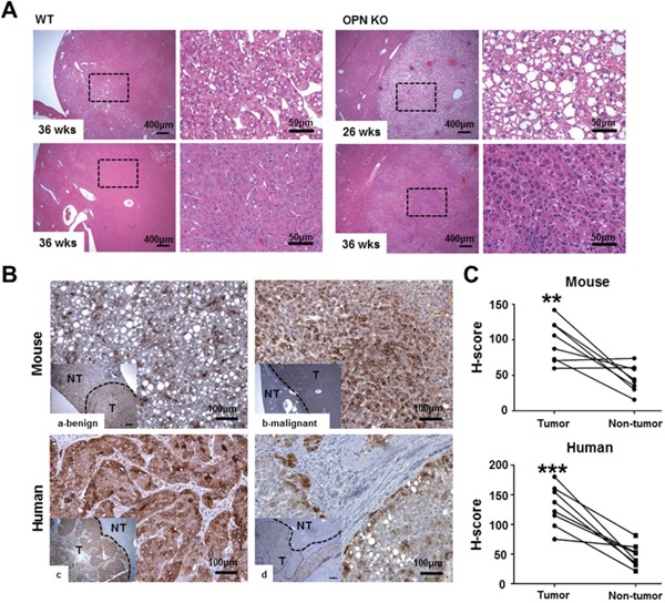
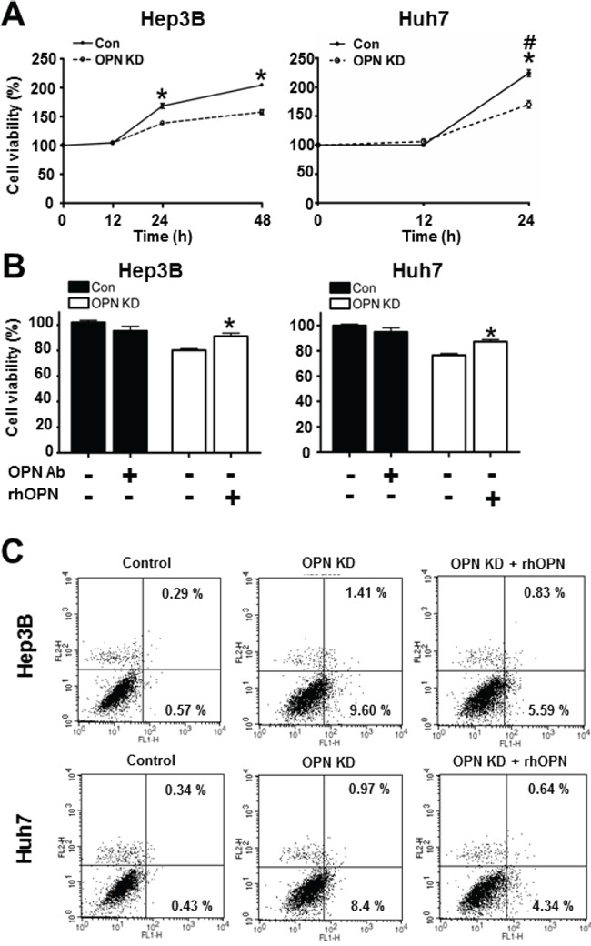
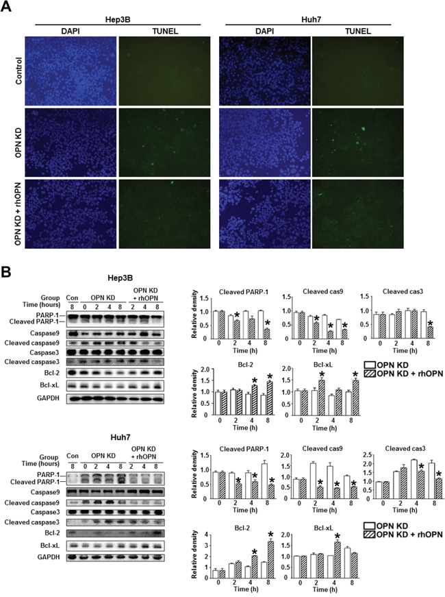
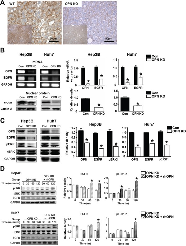
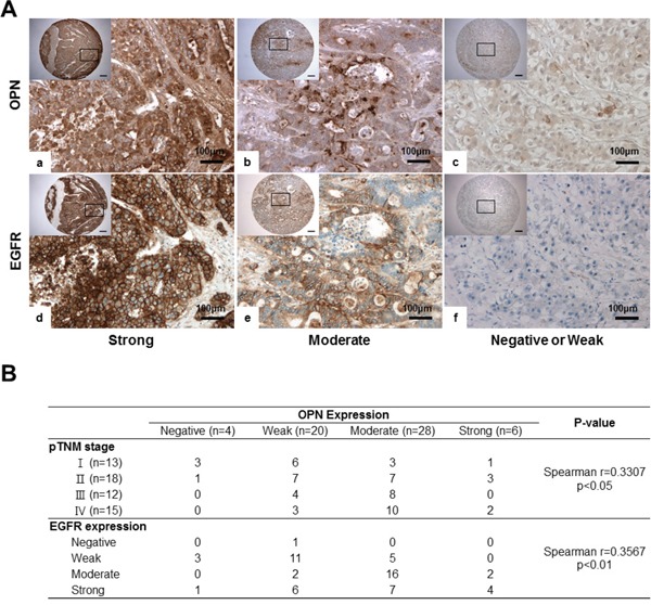
Previous clinical reports have found elevated osteopontin (OPN) levels in tumor tissues to be indicative of greater malignancy in human hepatocellular carcinoma (HCC). However, the role of OPN on carcinogenesis and its underlying mechanism remain unclear. In the present study, we investigated the oncogenic role of OPN in diethylnitrosamine (DEN)-induced hepatic carcinogenesis in mice. The overall incidence of hepatic tumors at 36 weeks was significantly lower in OPN knockout (KO) mice than in wild-type (WT) mice. Apoptosis was significantly enhanced in OPN KO mice, and was accompanied by the downregulation of epidermal growth factor receptor (EGFR). In the in vitro study, OPN suppression also led to lower mRNA and protein levels of EGFR associated with the downregulation of c-Jun in Hep3B and Huh7 human HCC cells lines, which resulted in increased apoptotic cell death in both cell lines. Moreover, a positive correlation was clearly identified between the expression of OPN and EGFR in human HCC tissues. These data demonstrate that the OPN deficiency reduced the incidence of chemically induced HCC by suppressing EGFR-mediated anti-apoptotic signaling. An important implication of our findings is that OPN positively contributes to hepatic carcinogenesis.

Keywords: EGFR; apoptosis; c-Jun; hepatocellular carcinoma; osteopontin.


Conflict of interest statement

CONFLICTS OF INTEREST

There are no conflicts of interest.

Figures

Figure 1
Figure 1. Histopathology and IHC for OPN
A. Hematoxylin and eosin staining in liver tissue samples from tumor-bearing WT and OPN KO mice. The boxed regions of the left panels in the WT and OPN KO samples are shown at higher magnification in the right panels. The nodules from the OPN KO mice at 26 and 36 weeks were sharply circumscribed and composed of well-differentiated tumor cells or vacuolated cells that formed trabeculae or nests, whereas the nodules from the WT mice at 36 weeks showed a sessile and solid growth pattern. B, C. IHC and quantification for OPN in mouse and human liver tissue samples. The tumor tissue samples (T) showed greater cytoplasmic expression of OPN than non-tumor tissue sample (NT) in both mouse (a, b) and human (c, d) samples. In mice, hepatocellular carcinoma (b) displayed more prominent OPN expression than hepatocellular adenoma (a). **P < 0.01 or ***P < 0.001 versus non-tumor tissue samples.
Figure 2
Figure 2. Apoptotic cell death in mouse liver tissue samples
A. Representative photomicrographs of total cells and TUNEL-positive cells in the liver tissue of DEN-treated-WT and OPN KO mice. The apoptotic index in non-tumor tissue of OPN KO mice at 26 weeks was considerably higher than that observed in WT mice. At 36 weeks, OPN KO mice also showed significantly larger numbers of apoptotic cells in non-tumor and tumor tissue samples than WT mice. B. Results are presented as means ± SEMs (n=3–4 for non-tumor tissue from WT and OPN KO mice, n=3–4 for tumor tissue from WT mice and n=2 for tumor tissue from OPN KO mice). **P < 0.01 versus WT mice.
Figure 3
Figure 3. Effect of OPN downregulation on cell viability
A. The cell viability of Hep3B and Huh7 at each time point. OPN KD Hep3B and Huh7 at 24 hours showed lower cell viability than control cells, and the cell viability of Hep3B was also decreased by OPN suppression at 48 hours. # The cell viability of Huh7 at 48 hours was not assessed due to saturation. Results are presented as SEMs (n=6 for each time point, three independent experiments). B. Changes in cell viability according to treatment with OPN antibody (Ab) or recombinant human OPN (rhOPN). The blockade of secreted OPN by OPN Ab in control cells caused a decrease of cell viability, although statistical significance was not observed. Supplemental rhOPN in OPN KD Hep3B and Huh7 considerably increased cell viability in both cell lines. The results are presented as means ± SEMs (n=6 for each condition, three independent experiments). C. Assessment of apoptotic cell death. Early and late apoptosis (lower and upper right quadrants) were more frequently observed in OPN KD Hep3B and Huh7. Supplemental rhOPN in OPN KD Hep3B and Huh7 caused a decrease in apoptosis.
Figure 4
Figure 4. Changes in apoptosis in relation to OPN suppression
A. TUNEL assay of the Hep3B and Huh7. OPN KD Hep3B and Huh7 showed a higher frequency of TUNEL-positive cells than control cells, and rhOPN treatment of OPN KD Hep3B and Huh7 reduced apoptotic cell death. B. Western blotting for the pro-apoptotic proteins PARP-1, caspase 9, and caspase 3, and the anti-apoptotic proteins Bcl-2 and Bcl-xL. The relative expression levels of apoptosis-related proteins in Hep3B (upper panels) and Huh7 (lower panels). The cleavage of PARP-1, caspase 9, and caspase 3 in OPN KD Hep3B and Huh7 from both cell lines was suppressed by supplementation with rhOPN, while the expression of anti-apoptotic proteins was upregulated. The results are presented as means ± SEMs (n=3 for each condition). * P < 0.05 versus OPN KD Hep3B and Huh7 under the corresponding culture conditions.
Figure 5
Figure 5. The regulatory effect of OPN on EGFR expression
A. Representative photomicrographs of immunohistochemistry for EGFR in liver tissue samples of WT and OPN KO mice at 36 weeks after DEN injection. EGFR expression in the cellular membrane of hepatocytes was stronger in WT mice than in OPN KO mice. Bar = 400 μm (inserts) B. RT-PCR and western blot for EGFR and c-Jun. OPN suppression caused a decrease in EGFR transcription, accompanied by the downregulation of nuclear c-Jun expression in the Hep3B and Huh7. The results are presented as SEMs (n=4). * P < 0.05 versus control cells. C. Western blot for EGFR and ERK. OPN KD cells Hep3B and Huh7 showed lower expression levels of EGFR and phosphorylated ERK1. The results are presented as means ± SEMs (n=4). * P < 0.05 versus control Hep3B and Huh7. D. Western blot for EGFR and ERK according to supplementation with rhOPN in OPN KD Hep3B and Huh7. The expression of EGFR and phosphorylated ERK was increased by rhOPN treatment in a time-dependent manner. The results are presented as means ± SEMs (n=3 for each condition). * P < 0.05 versus OPN KD Hep3B and Huh7.
Figure 6
Figure 6. IHC for OPN and EGFR in human HCC tissue samples
A. Representative photomicrographs of IHC for OPN and EGFR. Serially sectioned human HCC samples showing strong (a, d), moderate (b, e) and weak expression of OPN and EGFR (c, f). Bar = 400 μm (inserts). B. Correlations among OPN expression, pTNM, stages and EGFR expression.

References

    1. Ferlay J, Shin HR, Bray F, Forman D, Mathers C, Parkin DM. GLOBOCAN 2012 v1.0, Cancer Incidence and Mortality Worldwide: IARC CancerBase No. 2013;11
    1. Davila JA, Morgan RO, Shaib Y, McGlynn KA, El-Seraq HB. Hepatitis C infection and the increasing incidence of hepatocellular carcinoma: a population-based study. Gastroenterology. 2004;127:1372–1380. - PubMed
    1. Yang JD, Harmsen WS, Slettedahl SW, Chaiteerakij R, Enders FT, Therneau TM, Orsini L, Kim WR, Roberts LR. Factor that affect risk for hepatocellular carcinoma and effects of surveillance. Clin Gastroenterol Hepatol. 2011;9:617–623. - PubMed
    1. Baffy G, Brunt EM, Caldwell SH. Hepatocellular carcinoma in non-alcoholic fatty liver disease: A emerging menace. J Hepatol. 2012;56:1384–1391. - PubMed
    1. Ringelhan M, Protzer U. Oncogenic potential of hepatitis B virus encoded proteins. Curr Opin Virol. 2015;14:109–115. - PubMed