. 2016 Dec 27;7(52):86803-86815.
doi: 10.18632/oncotarget.13466.

Niclosamide and its analogs are potent inhibitors of Wnt/β-catenin, mTOR and STAT3 signaling in ovarian cancer

Affiliations

Niclosamide and its analogs are potent inhibitors of Wnt/β-catenin, mTOR and STAT3 signaling in ovarian cancer

Rebecca C Arend et al. Oncotarget. .

Erratum in

Abstract

Epithelial ovarian cancer (EOC) is the leading cause of gynecologic cancer mortality worldwide. Platinum-based therapy is the standard first line treatment and while most patients initially respond, resistance to chemotherapy usually arises. Major signaling pathways frequently upregulated in chemoresistant cells and important in the maintenance of cancer stem cells (CSCs) include Wnt/β-catenin, mTOR, and STAT3. The major objective of our study was to investigate the treatment of ovarian cancer with targeted agents that inhibit these three pathways. Here we demonstrate that niclosamide, a salicylamide derivative, and two synthetically manufactured niclosamide analogs (analog 11 and 32) caused significant inhibition of proliferation of two chemoresistant ovarian cancer cell lines (A2780cp20 and SKOV3Trip2), tumorspheres isolated from the ascites of EOC patients, and cells from a chemoresistant patient-derived xenograft (PDX). This work shows that all three agents significantly decreased the expression of proteins in the Wnt/β-catenin, mTOR and STAT3 pathways and preferentially targeted cells that expressed the ovarian CSC surface protein CD133. It also illustrates the potential of drug repurposing for chemoresistant EOC and can serve as a basis for pathway-oriented in vivo studies.

Keywords: Wnt pathway; cancer stem cells; chemoresistance; ovarian cancer; targeted therapy.


Conflict of interest statement

CONFLICTS OF INTERESTS

Authors declare no potential conflicts of interests.

Figures

Figure 1
Figure 1. Anti-proliferative effects of niclosamide, and its analogs, as single agents and in combination with chemotherapy and expression of CD133 post treatment
A. A2780ip2, A2780cp20, SKOV3ip1, SKOV3TRip2 cancer cell lines were treated with niclosamide, analog 11 or 32 (0.1- 4 μM) for 48 h. Level of ATP in the treated cells compared to the untreated cells were analyzed using ATPlite assay. B. A2780cp20 cells were treated concurrently with niclosamide or analogs in combination with carboplatin at indicated concentrations for 48 h. A combination index (CI) was calculated where CI <1 is synergistic. C. A2780cp20, SKOV3TRip2 cell lines were plated in 12 well plates and treated with niclosamide at indicated concentrations. Cell viability was measured by trypan blue exclusion method. All experiments were repeated 3 times. D. A2780cp20 cells were treated with niclosamide or analogs (2 μM), or IC50 doses of chemotherapy for 48 h and analyzed for CD133 expression by flow cytometry. Using student's t-test, *P < .05. All experiments were repeated 3 times. Data are represented as mean ± SD. Statistical analyses were performed using one-way ANOVA with application of Tukey's post test, P < .05 for all figures in (B).
Figure 2
Figure 2. Wnt/β-Catenin specific inhibition of ovarian cancer cell lines
A. A2780ip2, A2780cp20, SKOV3ip1 and SKOV3TRip2 cells in 24 well plates were treated with niclosamide and or Wnt3A, along with TOPflash construct and β-galactosidase-expressing vector in each well. After being incubated for 24 h, cells were analyzed for Wnt signaling. B. A2780ip2 and A2780cp20 cells were treated in 6 well plates with niclosamide and analogs at indicated concentrations for 24 h. The levels of LRP6 and phospo-LRP6, total β-catenin, were examined by western blot. All experiments were repeated 3 times. Data are represented as mean ± SD. Statistical analyses were performed by using student's t-tests, * P <0.05 when niclosamide group was compared to untreated control and niclosamide with Wnt3A group was compared to Wnt3A alone.
Figure 3
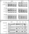
Figure 3. mTOR/STAT3 inhibition of ovarian cancer cell lines and patient samples
A. A2780ip2, A2780cp20 cell lines were plated in 6 well plates and were treated with niclosamide and analogs 11 and 32 at indicated concentrations for 24 h. Treated lysates were examined for the levels of mTOR pathway proteins (4E-BP1, phospho-4E-BP1, p(Thr389)-P70-70S6K, p70S6K). B. Acites cells from ovarian cancer patients were plated in 6 well plates and were treated with niclosamide at indicated concentrations for 24 h. Treated lysates were examined for the levels of STAT3 pathway proteins (p(Tyr705)-STAT3 and STAT3) and mTOR pathway proteins (p(Thr389)-p70-70SK, p70S6K, p(Ser235/236)-S6, and S6).
Figure 4
Figure 4. Cell cycle arrest by niclosamide and effect of niclosamide on cancer cell apoptosis and proliferation
A. A2780cp20 cell line was plated in 12 well plates and treated with indicated concentrations of niclosamide. Cells were stained with PI as described in Materials and Methods. Percentages of cells in different phases of the cell cycle were determined by flow cytometry at 24 and 48 h. B. A2780ip2, A2780cp20, SKOV3ip1, SKOV3TRip2 cancer cell lines were treated with niclosamide at indicated concentrations for 48 h. Floating and attached cells were combined for apoptosis detection by Cell Death ELISA kit for histone–associated DNA fragments as described in Materials and Methods. C. A2780ip2, A2780cp20 cell lines were treated with indicated concentrations of niclosamide. Lysates from treated cell lines were analyzed for survivin and cyclin D1. Data are represented as mean ± SD. Statistical analyses were performed by using student's t-tests, *P<.05, **P<.005 for figures (A) and (B).
Figure 5
Figure 5. Anti-proliferative effect of niclosamide and analogs 11 and 32 on PDX mouse model cells
PDX models 127 R (resistant) and 127 S (sensitive) were dissociated to single cell suspension and plated in 96 well plates. A. PDX cells were plated in tissue culture treated plates in 10% FBS + DMEM media. B. PDX cells were plated in low attachment plates with serum free x-vivo media with added supplements to promote stem cell growth. Cells were treated with niclosamide or analogs at increasing concentrations. Viability of cells was analyzed by ATPlite assay. All experiments were repeated 3 times. Data are represented as mean ± SD.

References

    1. Siegel RL, Miller KD, Jemal A. Cancer statistics, 2015. CA Cancer J Clin. 2015;65:5–29. doi: 10.3322/caac.21254. - DOI - PubMed
    1. Ricci F, Bernasconi S, Perego P, Ganzinelli M, Russo G, Bono F, Mangioni C, Fruscio R, Signorelli M, Broggini M, Damia G. Ovarian carcinoma tumor-initiating cells have a mesenchymal phenotype. Cell Cycle. 2012;11:1966–76. doi: 10.4161/cc.20308. - DOI - PubMed
    1. Takebe N, Harris PJ, Warren RQ, Ivy SP. Targeting cancer stem cells by inhibiting Wnt, Notch, and Hedgehog pathways. Nat Rev Clin Oncol. 2011;8:97–106. doi: 10.1038/nrclinonc.2010.196. - DOI - PubMed
    1. Zhou J, Wulfkuhle J, Zhang H, Gu P, Yang Y, Deng J, Margolick JB, Liotta LA, Petricoin E, 3rd, Zhang Y. Activation of the PTEN/mTOR/STAT3 pathway in breast cancer stem-like cells is required for viability and maintenance. Proc Natl Acad Sci U S A. 2007;104:16158–63. doi: 10.1073/pnas.0702596104. - DOI - PMC - PubMed
    1. Tomao F, Papa A, Strudel M, Rossi L, Lo Russo G, Benedetti Panici P, Ciabatta FR, Tomao S. Investigating molecular profiles of ovarian cancer: an update on cancer stem cells. J Cancer. 2014;5:301–10. doi: 10.7150/jca.8610. - DOI - PMC - PubMed

MeSH terms