A polysaccharide from Lentinus edodes inhibits human colon cancer cell proliferation and suppresses tumor growth in athymic nude mice
- PMID: 27888812
- PMCID: PMC5352182
- DOI: 10.18632/oncotarget.13481
A polysaccharide from Lentinus edodes inhibits human colon cancer cell proliferation and suppresses tumor growth in athymic nude mice
Abstract
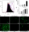
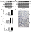
The antitumor effect of Lentinan is thought rely on the activation of immune responses; however, little is known about whether Lentinan also directly attacks cancer cells. We therefore investigated the direct antitumor activity of SLNT (a water-extracted polysaccharide from Lentinus edodes) and its probable mechanism. We showed that SLNT significantly inhibited proliferation of HT-29 colon cancer cells and suppressed tumor growth in nude mice. Annxein V-FITC/PI, DAPI, AO/EB and H&E staining assays all showed that SLNT induced cell apoptosis both in vitro and in vivo. SLNT induced apoptosis by activating Caspase-3 via both intrinsic and extrinsic pathways, which presented as the activation of Caspases-9 and -8, upregulation of cytochrome c and the Bax/Bcl-2 ratio, downregulation of NF-κB, and overproduction of ROS and TNF-α in vitro and in vivo. Pretreatment with the caspase-3 inhibitor Ac-DEVD-CHO or antioxidant NAC blocked SLNT-induced apoptosis. These findings suggest that SLNT exerts direct antitumor effects by inducing cell apoptosis via ROS-mediated intrinsic and TNF-α-mediated extrinsic pathways. SLNT may thus represent a useful candidate for colon cancer prevention and treatment.
Keywords: ROS; apoptosis; colon cancer; lentinan; nude mice.
Conflict of interest statement
The authors declare that they have no conflicts of interest.
Figures


Similar articles
-
A Lentinus edodes polysaccharide induces mitochondrial-mediated apoptosis in human cervical carcinoma HeLa cells.Int J Biol Macromol. 2017 Oct;103:676-682. doi: 10.1016/j.ijbiomac.2017.05.085. Epub 2017 May 17. Int J Biol Macromol. 2017. PMID: 28528001
-
Anti-tumor effect of polysaccharide from Hirsutella sinensis on human non-small cell lung cancer and nude mice through intrinsic mitochondrial pathway.Int J Biol Macromol. 2017 Jun;99:258-264. doi: 10.1016/j.ijbiomac.2017.02.071. Epub 2017 Feb 22. Int J Biol Macromol. 2017. PMID: 28235606
-
Sulfated lentinan induced mitochondrial dysfunction leads to programmed cell death of tobacco BY-2 cells.Pestic Biochem Physiol. 2017 Apr;137:27-35. doi: 10.1016/j.pestbp.2016.09.004. Epub 2016 Sep 24. Pestic Biochem Physiol. 2017. PMID: 28364801
-
Recent advances in polysaccharides from Lentinus edodes (Berk.): Isolation, structures and bioactivities.Food Chem. 2021 Oct 1;358:129883. doi: 10.1016/j.foodchem.2021.129883. Epub 2021 Apr 20. Food Chem. 2021. PMID: 33940295 Review.
-
Polysaccharides in Lentinus edodes: isolation, structure, immunomodulating activity and future prospective.Crit Rev Food Sci Nutr. 2014;54(4):474-87. doi: 10.1080/10408398.2011.587616. Crit Rev Food Sci Nutr. 2014. PMID: 24236998 Review.
Cited by
-
Properties of Yogurts Enriched with Crude Polysaccharides Extracted from Pleurotus ostreatus Cultivated Mushroom.Foods. 2023 Nov 5;12(21):4033. doi: 10.3390/foods12214033. Foods. 2023. PMID: 37959152 Free PMC article.
-
In Vitro Immuno-Modulatory Potentials of Purslane (Portulaca oleracea L.) Polysaccharides with a Chemical Selenylation.Foods. 2021 Dec 21;11(1):14. doi: 10.3390/foods11010014. Foods. 2021. PMID: 35010140 Free PMC article.
-
Simultaneous determination of spirodiclofen, spiromesifen, and spirotetramat and their relevant metabolites in edible fungi using ultra-performance liquid chromatography/tandem mass spectrometry.Sci Rep. 2021 Jan 15;11(1):1547. doi: 10.1038/s41598-021-81013-0. Sci Rep. 2021. PMID: 33452378 Free PMC article.
-
Stimulators of immunogenic cell death for cancer therapy: focusing on natural compounds.Cancer Cell Int. 2023 Sep 13;23(1):200. doi: 10.1186/s12935-023-03058-7. Cancer Cell Int. 2023. PMID: 37705051 Free PMC article. Review.
-
Anti-Cancerous Potential of Polysaccharide Fractions Extracted from Peony Seed Dreg on Various Human Cancer Cell Lines Via Cell Cycle Arrest and Apoptosis.Front Pharmacol. 2017 Mar 3;8:102. doi: 10.3389/fphar.2017.00102. eCollection 2017. Front Pharmacol. 2017. PMID: 28316571 Free PMC article.
References
-
- Jemal A, Murray T, Ward E, Samuels A, Tiwari RC, Ghafoor A, Feuer EJ, Thun MJ. Cancer Statistics, 2005. CA-Cancer J Clin. 2005;55:10–30. - PubMed
-
- Lin J, Qiu M, Xu R, Sandra Dobs A. Comparison of survival and clinicopathologic features in colorectal cancer among African American, Caucasian, and Chinese patients treated in the United States: Results from the surveillance epidemiology and end results (SEER) database. Oncotarget. 2015;6:33935–43. doi: 10.18632/oncotarget.5223. - DOI - PMC - PubMed
-
- Goldberg RM, Sargent DJ, Morton RF, Fuchs CS, Ramanathan RK, Williamson SK, Findlay BP, Pitot HC, Alberts SR. A randomized controlled trial of fluorouracil plus leucovorin, irinotecan, and oxaliplatin combinations in patients with previously untreated metastatic colorectal cancer. J Clin Oncol. 2004;22:23–30. - PubMed
-
- Chau I, Cunningham D. Adjuvant therapy in colon cancer—what, when and how? Ann Oncol. 2006;17:1347–1359. - PubMed
MeSH terms
Substances
LinkOut - more resources
Full Text Sources
Other Literature Sources
Molecular Biology Databases
Research Materials
Miscellaneous

