. 2016 Dec 1;167(6):1623-1635.e14.
doi: 10.1016/j.cell.2016.10.056. Epub 2016 Nov 23.

Structural Mechanism for Cargo Recognition by the Retromer Complex

Affiliations

Structural Mechanism for Cargo Recognition by the Retromer Complex

María Lucas et al. Cell. .

Abstract

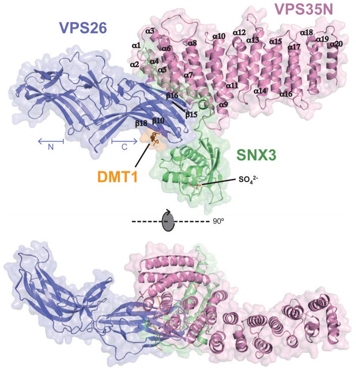
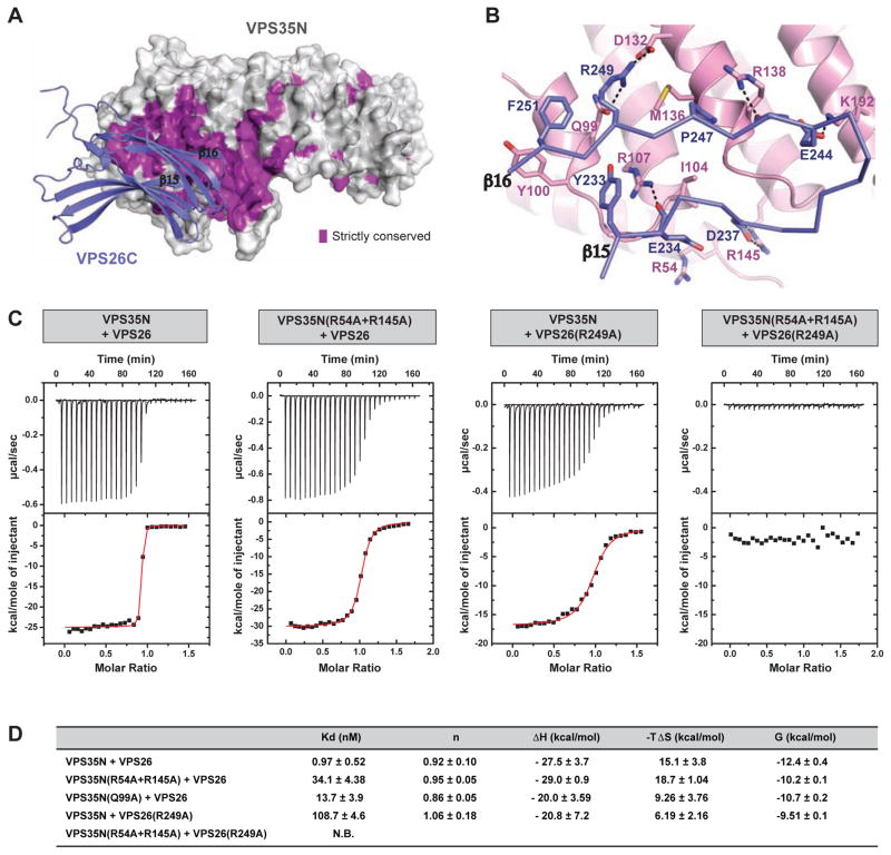
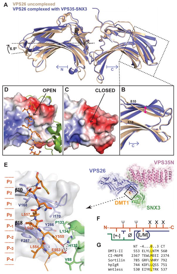
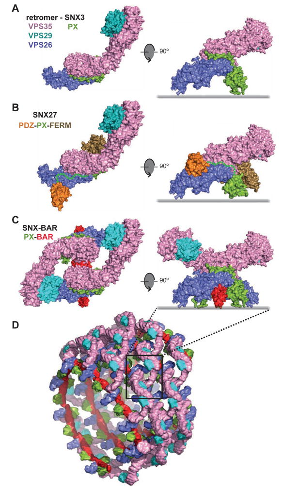
Retromer is a multi-protein complex that recycles transmembrane cargo from endosomes to the trans-Golgi network and the plasma membrane. Defects in retromer impair various cellular processes and underlie some forms of Alzheimer's disease and Parkinson's disease. Although retromer was discovered over 15 years ago, the mechanisms for cargo recognition and recruitment to endosomes have remained elusive. Here, we present an X-ray crystallographic analysis of a four-component complex comprising the VPS26 and VPS35 subunits of retromer, the sorting nexin SNX3, and a recycling signal from the divalent cation transporter DMT1-II. This analysis identifies a binding site for canonical recycling signals at the interface between VPS26 and SNX3. In addition, the structure highlights a network of cooperative interactions among the VPS subunits, SNX3, and cargo that couple signal-recognition to membrane recruitment.

Keywords: cargo recognition; endocytic recycling; endosomes; membrane recruitment; membrane tubules; protein coats; retrograde transport; retromer; sorting nexins; sorting signals.


Figures

Figure 1
Figure 1. Overall structure of the VPS26-VPS35N-SNX3-DMT1-II complex
The crystal structure is shown in two orthogonal views represented by a ribbon diagram with transparent surface. In the top view, the 20 α-helices (α1–20) that make up the solenoid of VPS35N and four β-strands from the C-terminal (CT) domain of VPS26 are labeled. Two sulphate ions (SO42−) found in the crystal structure, in stick representation, indicate the PtdIns3P-binding pocket on SNX3. See also Figure S1, Table S1 and Movie S1.
Figure 2
Figure 2. Interacting surfaces between VPS26 and VPS35
(A) Distribution of strictly conserved surface residues (violet) on VPS35N at the VPS26 contact site. (B) Relevant contacts of the VPS26-VPS35 interface. (C) Validation of the VPS26-VPS35 complex formation using ITC and site-directed mutagenesis. Baseline-corrected instrument response (upper) and integrated isotherms with the best fit curve to the data in red (lower) from ITC experiments measuring binding of VPS26 to VPS35N. (D) Thermodynamic binding parameters from ITC measurements. All ITC values are given as mean ± SD from at least three independent measurements. N.B., no appreciable binding. See also Figure S2
Figure 3
Figure 3. Structure of the retromer complex in solution
(A) SEC-MALS profiles for retromer at two protein concentrations under low and high ionic strength conditions. The value for the fitted molecular mass is shown as lines across the elution peak for each species. The predicted molecular mass of monomeric retromer is 150 kDa. (B) Normalized pair distance distribution P(r) functions for the monomeric and dimeric species of retromer. (C,D) Experimental spectrum of the small angle scattering of the monomeric (blue, C) and dimeric (red, D) species of retromer, and the simulated fit (grey) obtained from the model. (E,F) The ab initio shape reconstruction of the retromer complex by DAMMIN using P1 symmetry for the monomer (E) and P2 for the dimer (F), showing the fit with the crystallographic structures of VPS26-VPS35N and VPS26-VPS35C (PDB codes: 5F0L and 2R17). (G) Retromer dimer bound to two SNX3 molecules sits parallel to the membrane plane. Positively-charged VPS26 N-lobe provides a complementary surface for membrane interaction. See also Figure S3 and Movie S2.
Figure 4
Figure 4. Close-up of the SNX3 interfaces for retromer recruitment to membranes
(A) SNX3 PX domain oriented to show the two sulphate ions at the phosphoinositide-binding pocket and superimposed with the Grd19p PX domain bound to C4-PtdIns(3)P. (B) Contacts between the N-terminal tail of SNX3 and the VPS26-VPS35 subcomplex. (C) Contacts between β1 of SNX3 and the α8-α9 connecting loop of VPS35. (D) Close-up view of the SNX3 P133 insertion between strands β10 and β18 of VPS26. See also Figure S4.
Figure 5
Figure 5. Structural plasticity of VPS26 for cargo recognition
(A) Superimposition of the crystal structures of VPS26A, in the free (PDB: 2FAU, brown) and VPS26-VPS35N-SNX3-DMT1-II complexed form (current work, PDB: 5F0L, blue). (B) Close-up view of the C-terminal domain. Pink arrow indicates changes in strand β10 from basal to active state. (C,D) Same view as in (B) showing the electrostatic surface potential (ranging from blue 5 kTe−1 to red - 5 kTe−1) of basal VPS26A (C) and activated VPS26A bound to the recycling signal of DMT1-II (D). (E) Close-up view showing the recognition of the DMT1-II recycling motif by the VPS26-SNX3 subcomplex. (F) Cartoon representing the consensus VPS26-SNX3 cargo binding motif (X stands for any residue, Ø a bulky aromatic residue, Ψ a residue having a hydrophobic or long aliphatic hydrocarbon tail, and [+-] any charged residue). (G) Sequence alignment of representative retromer-binding motifs. See also Figure S5 and Movie S3.
Figure 6
Figure 6. SNX3 recruits retromer to membranes, promoting DMT1-II recycling
(A) ITC isotherms of the binding of retromer to wild-type SNX3 and various SNX3 deletion/substitution mutants in the absence or presence of the DMT1-II recycling signal (residues 550–568). (B) ITC isotherms for the binding of peptides encompassing the normal DMT1-II recycling signal (residues 550–568) or a mutant of this sequence with Y555A and L557A substitutions (mut), to SNX3, retromer, or a combination of SNX3 with retromer or with retromer having VPS26 F287A and V168N substitutions in VPS26 (mut). (C) Immunoblot analysis of wild-type (WT) and SNX3-KO HeLa cells using antibodies to SNX3 and GADPH (loading control). The positions of molecular mass markers (in kDa) are indicated. (D) Immunofluorescence microscopy of endogenous VPS26 in WT, SNX3-KO, or SNX3-KO HeLa cells rescued (res) with GFP-SNX3 or GFP-SNX3(ΔN). Bars: 10 μm. (E) Quantification of the recruitment of endogenous VPS26 to membranes by different GFP-SNX3 constructs expressed in SNX3-KO cells. Datasets are from the stable cells lines shown in panel D, as well as stable cell lines expressing the GFP-SNX3(RRY), GFP-SNX3(HPL) or GFP-SNX3(ED) mutants shown in Figure S6F. Bars represent the mean ± SEM (n=22–34 cells; * p<0.05, *** p<0.005 by Student’s t-test). (F) Immunofluorescence microscopy of endogenous EEA1 and ectopically-expressed DMT1-II in the same cell lines from panel D. Bars: 10 μm. Magnified views of the boxed areas are shown on the right. Bars: 2 μm. Images in D and F are maximum intensity projections of Z-stacks. Nuclei were stained with DAPI (blue). (G) Quantification of DMT1-II–EEA1 colocalization in the cell lines shown in panel F and Figure S7. Bars represent the mean ± SEM of the Pearson’s correlation coefficient from three independent experiments (n=10–19; ns: not significant, *** p<0.005 by Student’s t-test). See also Figures S6 and S7.
Figure 7
Figure 7. Proposed architecture of different SNX-retromer assemblies
(A) Proposed model of the SNX3-retromer complex. The entire retromer (VPS26-VPS29-VPS35) structure was generated by fitting the crystal structures of VPS26-VPS35N (current work) and VPS29-VPS35C (Hierro et al., 2007) within experimental SAXS data (Figure 3E) and superimposed on the crystal structure of SNX3 (green) bound to VPS26-VPS35N. (B) Proposed model of the SNX27-retromer complex. The retromer (VPS26-VPS29-VPS35) structure was superimposed on the crystal structure of the SNX27 PDZ domain (orange) bound to VPS26 (Gallon et al., 2014). Residues for the linker segment between the PDZ and PX domains of SNX27 are indicated with a green dashed line. The PX domain (green) of SNX27 (PDB: 4HAS) was superimposed on the PX domain of SNX3 and linked to the crystal structure of the FERM-like domain of SNX17 (Ghai et al., 2013). (C) Proposed model of a SNX-BAR-retromer complex. Two VPS26-VPS29-VPS35 retromer complexes were superimposed on the SNX9 PX-BAR dimer structure (Pylypenko et al., 2007) using the PX-domain of SNX3 as reference. (D) Cartoon showing a speculative helical coat organization formed by the combination of retromer and PX-BAR dimers. See also Movie S4.

Comment in

References

    1. Adams PD, Afonine PV, Bunkoczi G, Chen VB, Davis IW, Echols N, Headd JJ, Hung LW, Kapral GJ, Grosse-Kunstleve RW, et al. PHENIX: a comprehensive Python-based system for macromolecular structure solution. Acta crystallographica Section D, Biological crystallography. 2010;66:213–221. - PMC - PubMed
    1. Arighi CN, Hartnell LM, Aguilar RC, Haft CR, Bonifacino JS. Role of the mammalian retromer in sorting of the cation-independent mannose 6-phosphate receptor. The Journal of cell biology. 2004;165:123–133. - PMC - PubMed
    1. Canuel M, Lefrancois S, Zeng J, Morales CR. AP-1 and retromer play opposite roles in the trafficking of sortilin between the Golgi apparatus and the lysosomes. Biochemical and biophysical research communications. 2008;366:724–730. - PubMed
    1. Carlton J, Bujny M, Peter BJ, Oorschot VM, Rutherford A, Mellor H, Klumperman J, McMahon HT, Cullen PJ. Sorting nexin-1 mediates tubular endosome-to-TGN transport through coincidence sensing of high-curvature membranes and 3-phosphoinositides. Current biology : CB. 2004;14:1791–1800. - PubMed
    1. Cereghino JL, Marcusson EG, Emr SD. The cytoplasmic tail domain of the vacuolar protein sorting receptor Vps10p and a subset of VPS gene products regulate receptor stability, function, and localization. Molecular biology of the cell. 1995;6:1089–1102. - PMC - PubMed

MeSH terms

Substances