. 2016 Dec;18(12):775-784.
doi: 10.1016/j.neo.2016.11.002. Epub 2016 Nov 25.

Mirvetuximab Soravtansine (IMGN853), a Folate Receptor Alpha-Targeting Antibody-Drug Conjugate, Potentiates the Activity of Standard of Care Therapeutics in Ovarian Cancer Models

Affiliations

Mirvetuximab Soravtansine (IMGN853), a Folate Receptor Alpha-Targeting Antibody-Drug Conjugate, Potentiates the Activity of Standard of Care Therapeutics in Ovarian Cancer Models

Jose F Ponte et al. Neoplasia. 2016 Dec.

Abstract

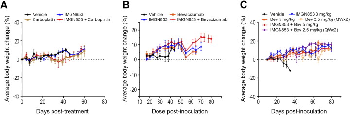
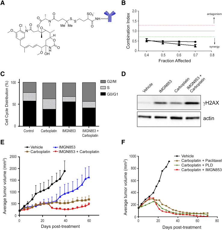
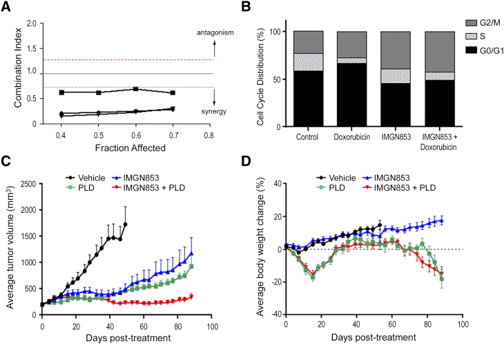
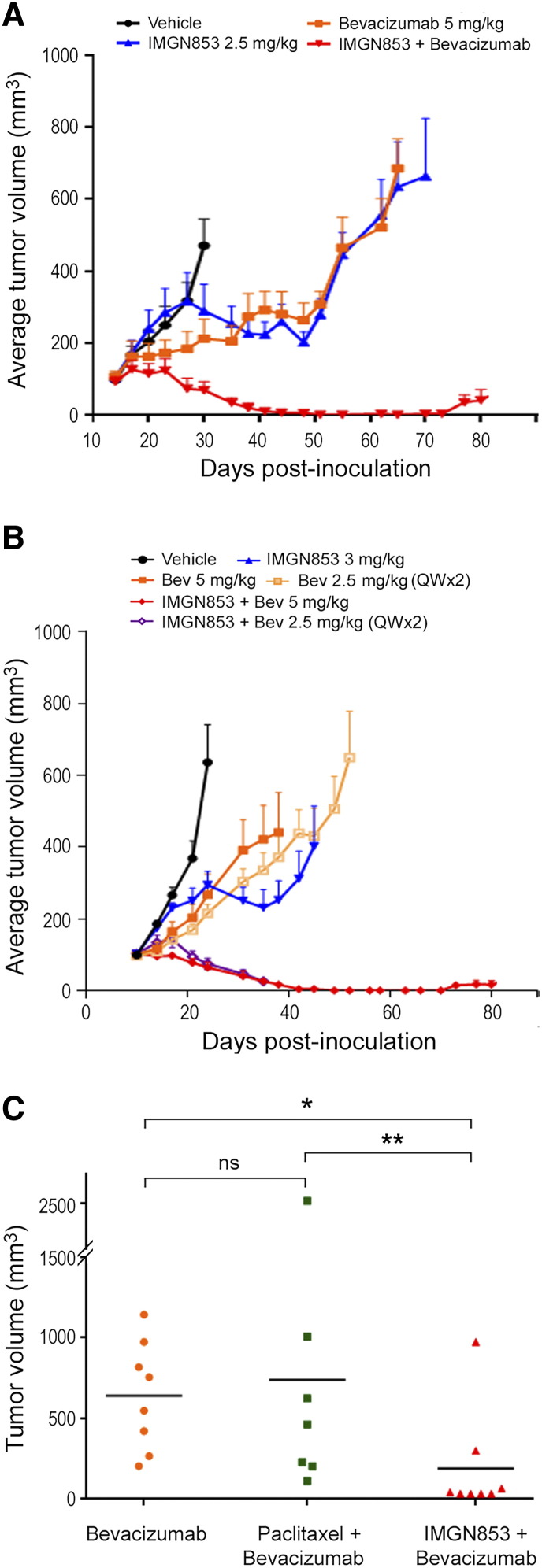
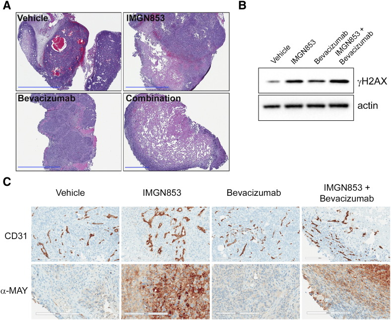
Elevated folate receptor alpha (FRα) expression is characteristic of epithelial ovarian cancer (EOC), thus establishing this receptor as a candidate target for the development of novel therapeutics to treat this disease. Mirvetuximab soravtansine (IMGN853) is an antibody-drug conjugate (ADC) that targets FRα for tumor-directed delivery of the maytansinoid DM4, a potent agent that induces mitotic arrest by suppressing microtubule dynamics. Here, combinations of IMGN853 with approved therapeutics were evaluated in preclinical models of EOC. Combinations of IMGN853 with carboplatin or doxorubicin resulted in synergistic antiproliferative effects in the IGROV-1 ovarian cancer cell line in vitro. IMGN853 potentiated the cytotoxic activity of carboplatin via growth arrest and augmented DNA damage; cell cycle perturbations were also observed in cells treated with the IMGN853/doxorubicin combination. These benefits translated into improved antitumor activity in patient-derived xenograft models in vivo in both the platinum-sensitive (IMGN853/carboplatin) and platinum-resistant (IMGN853/pegylated liposomal doxorubicin) settings. IMGN853 co-treatment also improved the in vivo efficacy of bevacizumab in platinum-resistant EOC models, with combination regimens causing significant regressions and complete responses in the majority of tumor-bearing mice. Histological analysis of OV-90 ovarian xenograft tumors revealed that concurrent administration of IMGN853 and bevacizumab caused rapid disruption of tumor microvasculature and extensive necrosis, underscoring the superior bioactivity profile of the combination regimen. Overall, these demonstrations of combinatorial benefit conferred by the addition of the first FRα-targeting ADC to established therapies provide a compelling framework for the potential application of IMGN853 in the treatment of patients with advanced ovarian cancer.


Figures

Supplementary Figure S1
Body weight changes in multiple EOC xenograft models. (A) Mice bearing platinum-sensitive ovarian cancer PDXs were dosed with a single administration of IMGN853 (2.5 mg/kg) or carboplatin (80 mg/kg), alone and in combination. Body weights were measured twice weekly. Mean values are plotted against vehicle controls. (B) Mice bearing established OV-90 xenografts were dosed with a single administration of IMGN853 (2.5 mg/kg) or bevacizumab (5 mg/kg), alone and in combination. Body weights were measured twice weekly. Mean values are plotted against vehicle controls. (C) Mice bearing established OV-90 xenografts received a single injection of 3 mg/kg IMGN853 alone or in combination with bevacizumab, administered either as a single 5-mg/kg dose or two consecutive weekly doses of 2.5 mg/kg (QW × 2). Body weights were measured twice weekly. Mean values are plotted against vehicle controls.
Figure 1
Figure 1
IMGN853 potentiates the therapeutic activity of carboplatin in platinum-sensitive and platinum-resistant EOC models. (A) Structural representation of IMGN853. (B) IGROV-1 cells were treated with graded concentrations of IMGN853, carboplatin, or both, and the effects on proliferation were determined. The CI was calculated using median effect analysis. Data from two independent experiments are shown, determined for a range of drug concentrations and a fractional effect (Fa) of 0.4 to 0.7. Data points below the green line represent synergy between the drug pairs. (C) IGROV-1 cells were treated with carboplatin (20 μM) or IMGN853 (8 nM), both alone and in combination, for 6 hours. Cells were washed, and cell cycle distribution was determined following 24-hour culture in drug-free medium. (D) IGROV-1 cells were exposed to carboplatin (40 μM) or IMGN853 (16 nM), alone or in combination, for 6 hours followed by incubation in drug-free medium for an additional 18 hours. Cellular extracts were immunoblotted for γH2AX or actin (loading control) as indicated. (E) Platinum-sensitive ovarian cancer PDXs were established in SCID mice. Animals were dosed with a single administration of IMGN853 (2.5 mg/kg) or carboplatin (80 mg/kg), alone and in combination (n = 7 mice/group). Data are expressed as mean and SEM for each time point. (F) Mice bearing platinum-sensitive PDX tumors (n = 7 mice/group) received two consecutive weekly doses of vehicle, carboplatin (80 mg/kg, IP) plus paclitaxel (10 mg/kg), carboplatin plus PLD (4 mg/kg), or carboplatin plus IMGN853 (5 mg/kg), as indicated.
Figure 2
Figure 2
The anticancer activity of doxorubicin (in vitro) and PDL (in vivo) is enhanced by IMGN853. (A) IGROV-1 cells were treated with increasing concentrations of IMGN853, doxorubicin, or both, and the effects on proliferation were determined. The CI was calculated using median effect analysis. Data from three independent experiments are shown, with points below the green line representing synergy between the drug pair. (B) IGROV-1 cells were treated with doxorubicin (200 nM) or IMGN853 (8 nM), both alone and in combination, for 6 hours. Cells were washed, and cell cycle distribution was determined following 24-hour culture in drug-free medium. (C) Platinum-resistant ovarian cancer PDXs were established in SCID mice. Animals received two consecutive weekly doses of IMGN853 (5 mg/kg) and PLD (4 mg/kg) alone or in combination (n = 8 mice/group). Data are expressed as mean and SEM for each time point. (D) Body weights were measured twice weekly. Mean values are plotted against vehicle controls.
Figure 3
Figure 3
In vivo antitumor activity of IMGN853 in combination with bevacizumab. (A) Mice bearing established OV-90 xenografts (n = 6 mice/group) were IV dosed with a single administration of IMGN853 (2.5 mg/kg) or bevacizumab (5 mg/kg), alone and in combination, as indicated. Data are expressed as mean and SEM for each time point. The combination of IMGN853 and bevacizumab led to prolonged tumor regressions. (B) Mice bearing established OV-90 xenografts (n = 7 mice/group) received a single IV injection of IMGN853 (3 mg/kg) alone or in combination with bevacizumab, administered either as a single 5-mg/kg dose or two consecutive weekly doses of 2.5 mg/kg (QW × 2). Combination treatments of IMGN853 with either bevacizumab dosing regimen were more efficacious than the corresponding single agents. (C) Mice bearing platinum-resistant ovarian cancer PDXs received two consecutive weekly doses of bevacizumab (5 mg/kg), alone or in combination with either paclitaxel (10 mg/kg) or IMGN853 (5 mg/kg), and tumor growth was monitored out to 102 days. Tumor volumes were measured at the end of study, and individual tumor sizes were plotted according to treatment group. *P = .011; ** P = .018; ns, not significant (Wilcoxon test, nonadjusted).
Figure 4
Figure 4
Combination IMGN853 plus bevacizumab treatment disrupts tumor vascularization and causes rapid tumor destruction in OV-90 xenografts. (A) OV-90 tumor-bearing mice were treated with a single dose of vehicle, IMGN853 (2.5 mg/kg), bevacizumab (5 mg/kg), or IMGN853 plus bevacizumab, and tumors were harvested 4 days later. Histological staining (H&E) revealed the presence of large, central necrotic zones in tumors from combination-treated mice. Original magnification, 4×; scale bars, 2 mm (for combination panel, 600 μm). (B) Tumor extracts were immunoblotted for γH2AX or actin (loading control) as indicated. (C) Immunohistochemical evaluation of CD31 expression (upper panel) and maytansine detection (anti-MAY; lower panel) in tumor tissues on day 4. For each group, representative micrographs from one of three tumors are shown. Original magnification, 20×; scale bars, 200 μm.

References

    1. Siegel R., Ma J., Zou Z., Jemal A. Cancer statistics, 2014. CA Cancer J Clin. 2014;64:9–29. - PubMed
    1. Society AC . American Cancer Society; 2016. Cancer facts & figures.
    1. Lacey J.V., Sherman M.E. In: Ovarian neoplasia. Robboy S.L., Mutter G.L., Prat J., editors. vol. 2. Churchill Livingstone Elsevier; Oxford: 2009. p. 601.
    1. Liu J., Matulonis U.A. New strategies in ovarian cancer: translating the molecular complexity of ovarian cancer into treatment advances. Clin Cancer Res. 2014;20:5150–5156. - PubMed
    1. Luvero D., Milani A., Ledermann J.A. Treatment options in recurrent ovarian cancer: latest evidence and clinical potential. Ther Adv Med Oncol. 2014;6:229–239. - PMC - PubMed

MeSH terms