Review
. 2017 Jun:107:228-244.
doi: 10.1016/j.freeradbiomed.2016.11.039. Epub 2016 Nov 24.

Coordination of DNA single strand break repair

Affiliations
Review

Coordination of DNA single strand break repair

Rachel Abbotts et al. Free Radic Biol Med. 2017 Jun.

Abstract

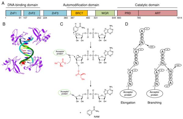
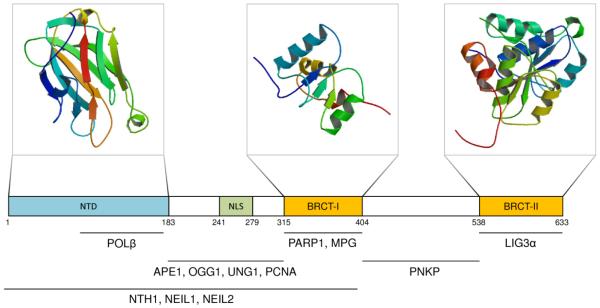
The genetic material of all organisms is susceptible to modification. In some instances, these changes are programmed, such as the formation of DNA double strand breaks during meiotic recombination to generate gamete variety or class switch recombination to create antibody diversity. However, in most cases, genomic damage is potentially harmful to the health of the organism, contributing to disease and aging by promoting deleterious cellular outcomes. A proportion of DNA modifications are caused by exogenous agents, both physical (namely ultraviolet sunlight and ionizing radiation) and chemical (such as benzopyrene, alkylating agents, platinum compounds and psoralens), which can produce numerous forms of DNA damage, including a range of "simple" and helix-distorting base lesions, abasic sites, crosslinks and various types of phosphodiester strand breaks. More significant in terms of frequency are endogenous mechanisms of modification, which include hydrolytic disintegration of DNA chemical bonds, attack by reactive oxygen species and other byproducts of normal cellular metabolism, or incomplete or necessary enzymatic reactions (such as topoisomerases or repair nucleases). Both exogenous and endogenous mechanisms are associated with a high risk of single strand breakage, either produced directly or generated as intermediates of DNA repair. This review will focus upon the creation, consequences and resolution of single strand breaks, with a particular focus on two major coordinating repair proteins: poly(ADP-ribose) polymerase 1 (PARP1) and X-ray repair cross-complementing protein 1 (XRCC1).

Keywords: Aging; DNA repair; Neurodegeneration; Oxidative DNA damage; PARP1; XRCC1.


Figures

Figure 1
Figure 1
Overview of BER. 1. BER is initiated by substrate-specific recognition and excision of a damaged base by a DNA glycosylase (e.g. OGG1), creating an abasic site. 2. APE1 incises the DNA backbone at the AP site, generating a strand break with 3′-hydroxyl and 5′-deoxyribosephosphate (dRP) flanking groups. Bifunctional glycosylases with AP lyase activity can also cleave the abasic site, generating a 3′-α,β-unsaturated aldehyde (or 3′-phosphate) and 5′-phosphate termini; blocking termini require additional processing (see text). 3. The excised nucleotide is replaced by DNA polymerase repair synthesis. A. Short-patch repair synthesis by POLβ involves the incorporation of a single nucleotide. B. In the long-patch pathway, a DNA polymerase (δ, ε or β) incorporates 2-12 nucleotides by strand displacement synthesis in conjunction with the sliding clamp PCNA, creating a flap intermediate that is removed by FEN1 (4). 5. The DNA nick is sealed by a ligase, namely DNA LIG3α (likely in complex with XRCC1) or LIG1. PARP1 preumably participates in the response when DNA damage or repair intermediates are not engaged by the classic BER machinery.
Figure 2
Figure 2
PARP1. A. PARP1 domain architecture. ZnF1-3: zinc finger domains 1-3; BRCT: BRCA1 C-terminus domain; WGR: tryptophan-, glycine- arginine-rich domain; PRD: PARP regulatory domain; ART: catalytic domain, highly-conserved in other ADP-ribosyl transferases. B. Crystal structure of PARP1 DBD in complex with DNA (PDB ID 4AV1) [79]. C. Schematic of ADP-ribosylation as catalyzed by PARP1, in which an ADP-ribose molecule is transferred from NAD+ to an acceptor protein with the release of nicotinamide (NAM). Red structures represent critical residues of the PARP1 active site. Nu: acceptor protein nucleophile. D. Elongation and branching reactions use the same chemistry, but vary in the orientation of the riboses. Elongation involves a 1′′ → 2′ ribose-ribose (A-ribose) bond. Branching (every ~20 ADP-ribose units) involves a 1′′′ → 2′ ribose-ribose glycosidic (N-ribose) bond.
Figure 3
Figure 3
XRCC1. XRCC1 domain architecture with crystal structures of the N-terminal domain (NTD; PDB ID 1XNA [166]), BRCT-I (PDB ID 1CDZ [165]) and BRCT-II (PDB ID 3PC6 [167]). Also indicated are sites of interaction with other BER and SSBR proteins; see text for abbreviations. NLS = nuclear localization sequence.
Figure 4
Figure 4
Obstructive 5′ and 3′ strand break termini and the repair enzymes required for resolution. PARP1 and XRCC1 regulate the stability, complex assembly and coordination, and/or enzymatic activity of many of these repair factors as described in the text. Enzymes in green are known to interact with XRCC1, enzymes in orange are known to interact with both XRCC1 and PARP1, and enzymes in blue are known to interact with XRCC1 and possess a prototypical PAR binding site that may mediate an interaction with PARP1. PNKP: polynucleotide kinase 3′-phosphatase; POLβ: DNA polymerase β; FEN1: flap endonuclease 1; APTX: aprataxin; APE1: apurinic/apyrimidinic endonuclease 1; TDP1: tyrosyl DNA phosphodiesterase 1.

References

    1. Lindahl T. Instability and decay of the primary structure of DNA. Nature. 1993;362(6422):709–15. - PubMed
    1. Olinski R, Jurgowiak M, Zaremba T. Uracil in DNA--its biological significance. Mutat Res. 2010;705(3):239–45. - PubMed
    1. De Bont R, van Larebeke N. Endogenous DNA damage in humans: a review of quantitative data. Mutagenesis. 2004;19(3):169–85. - PubMed
    1. Rydberg B, Lindahl T. Nonenzymatic methylation of DNA by the intracellular methyl group donor S-adenosyl-L-methionine is a potentially mutagenic reaction. EMBO J. 1982;1(2):211–6. - PMC - PubMed
    1. Gates KS, Nooner T, Dutta S. Biologically relevant chemical reactions of N7-alkylguanine residues in DNA. Chem Res Toxicol. 2004;17(7):839–56. - PubMed

Publication types

Substances