. 2017 Jul;31(7):1582-1592.
doi: 10.1038/leu.2016.354. Epub 2016 Nov 28.

Extended time-lapse in vivo imaging of tibia bone marrow to visualize dynamic hematopoietic stem cell engraftment

Affiliations

Extended time-lapse in vivo imaging of tibia bone marrow to visualize dynamic hematopoietic stem cell engraftment

S Kim et al. Leukemia. 2017 Jul.

Abstract

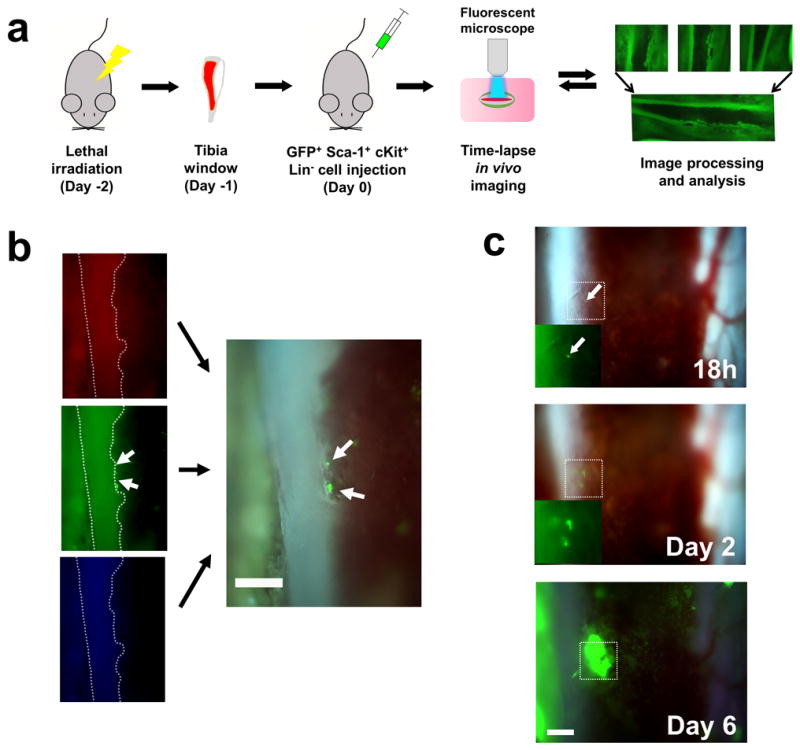
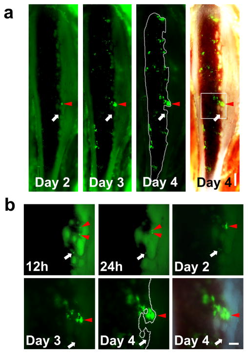
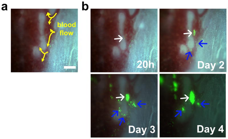
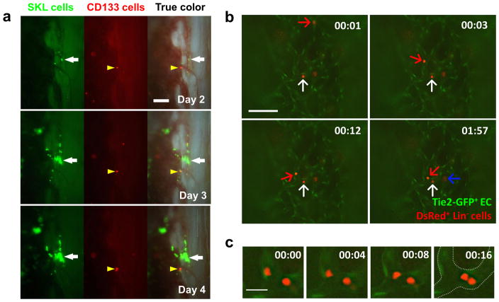
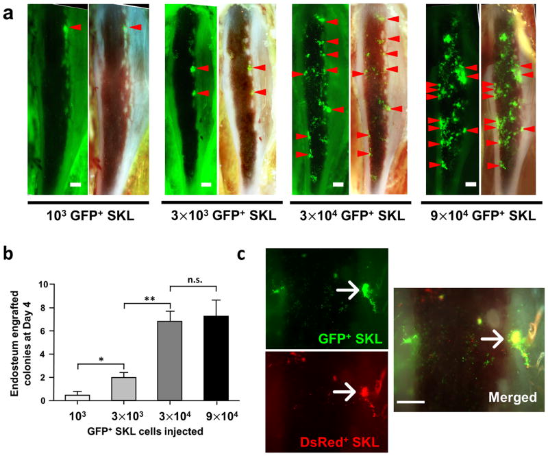
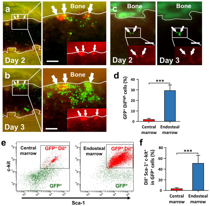
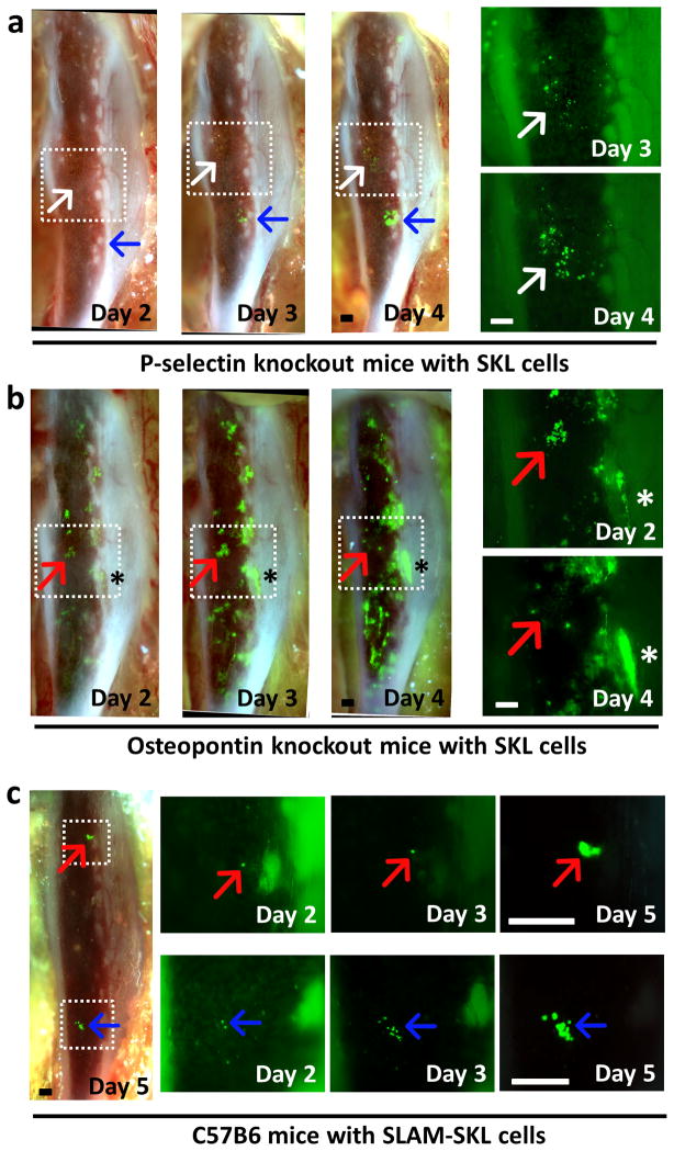
Homing, engraftment and proliferation of hematopoietic stem/progenitor cell (HSC/HPCs) are crucial steps required for success of a bone marrow transplant. Observation of these critical events is limited by the opaque nature of bone. Here we demonstrate how individual HSCs engraft in long bones by thinning one side of the tibia for direct and unbiased observation. Intravital imaging enabled detailed visualization of single Sca-1+, c-Kit+, Lineage- (SKL) cell migration to bone marrow niches and subsequent proliferation to reconstitute hematopoiesis. This longitudinal study allowed direct observation of dynamic HSC/HPC activities during engraftment in full color for up to 6 days in live recipients. Individual SKL cells, but not mature or committed progenitor cells, preferentially homed to a limited number of niches near highly vascularized endosteal regions, and clonally expanded. Engraftment of SKL cells in P-selectin and osteopontin knockout mice showed abnormal homing and expansion of SKL cells. CD150+, CD48- SKL populations initially engrafted in the central marrow region, utilizing only a subset of niches occupied by the parent SKL cells. Our study demonstrates that time-lapse imaging of tibia can be a valuable tool to understand the dynamic characteristics of functional HSC and niche components in various mouse models.


Conflict of interest statement

CONFLICT OF INTEREST

Authors declare no conflict of interest.

Figures

Figure 1
Figure 1. Time-lapse in vivo imaging of the tibia bone to visualize the engraftment of SKL cells
(a) A diagram showing the processes of in vivo tibia imaging. Mice were lethally irradiated 2 days before cell injection. Each image was tiled into a mosaic to create the panoramic view of the tibia marrow. (b) Use of the RGB filter for real-time, true color video recording. Color-separated images from RGB channel demonstrate that individual GFP+ cells could be clearly visualized using 10× magnification (white arrows). Non-fluorescent SKL cells from C57BL6 mice did not generate any signal in the recipients (data not shown). (c) Formation of BM colonies from individual SKL cell in the RGB channel. The fate of a single GFP+ SKL cell was followed over 6 days. All scale bars = 200μm.
Figure 2
Figure 2. In vivo imaging of the same mouse over time demonstrates that SKL cells tend to engraft and proliferate near the endosteal region
(a) Mosaic images of time-lapse in vivo imaging of a tibia window. 3×10⁴ GFP+ SKL cells were injected at Day 0 and in vivo colony formation from SKL cells was monitored in the same animal over time (n=6). Scale bar=500μm (b) Higher magnification of single GFP+ cells engrafted on the endosteal surface developing into a colony (the boxed region at Day 4). Injected SKL cells homed to the marrow and developed into a colony over 4 days (Red arrowheads, the same area marked at the mosaic images). However, not every cell homed to the endosteal surface formed colonies (white arrows). Scale bar=200μm.
Figure 3
Figure 3. Time-lapse imaging of the individual SKL cells showing endosteal engraftment
(a) Blood flow (arrows) in the endosteal region. (b) Time lapse imaging of the area showing the engraftment of individual SKL cells. The white arrow indicates the main colony and blue arrows indicate the satellite colonies developed at later time points. Scale bar=100μm.
Figure 4
Figure 4. Time-lapse and live in vivo imaging to visualize the engraftment process
(a) Time-lapse in vivo imaging of tibia co-transplanted with 3×10⁴ GFP+ SKL cells (white arrow) and 105 DsRed+ CD133+ hematopoietic progenitor cells (yellow arrowhead, n=3). (b) 5×10⁶ DsRed+ Lin cells were injected to Tie2-GFP mice to visualize the dynamic interaction of Tie2-GFP+ BM endothelium (EC) and engrafting cells (n=5). Images were simultaneously captured from Video S4 showing rolling/tethering (red arrow) and attaching/extravasating cells (white arrow) that migrate into BM cavity. A cell that is already crossed the endothelial barrier was also observed (blue arrow). Scale bar=200 μm. The observation time is same as appeared in Video S5. (c) Higher magnification images showing cells that tether and scan along the endothelium via millipede-like locomotion. Scale bar = 20μm. Time format = (mm:ss).
Figure 5
Figure 5. SKL cells tend to engraft on the specific niche with the limited number
(a) Representative images of tibia window with different number of GFP+ SKL cell injection at day 4. Endosteum-engrafted colonies above 3500μm2 were marked with red arrowheads to speculate possible osteoblastic niche locations. (b) The average number of observed colonies in each group was shown in graph (n=5–10). Data analyzed by ANOVA and Tukey’s post test, *, P<0.05; **, P<0.01; n.s.= not significant (c) The same number of SKL cells from GFP and DsRed mice (3×10⁴ cells) were injected into the same recipient to show the niche preference of SKL cells (n=3). Scale bars = 500μm for a, 300μm for c.
Figure 6
Figure 6. Dye retaining cells are located on the endosteal region
(a and b) Time-lapse in vivo tracking of dye retaining cells to visualize slow cycling cells (n=5). 4×10³ GFP+ SKL cells stained with DiI membrane-dye (red) were injected into the femoral artery and monitored for engraftment. GFP+ DiI bright cells were located predominantly near the endosteum (white arrows). (c) The DiI dye that was initially observed in GFP+ SKL cells at the central marrow region at day 2 was rapidly diluted after cell proliferation after 24h. (d–f) FACS analysis of the dye retaining cells from the tibia at day 3 (n=5). Student’s t-test was done for statistical analysis. ***, P<0.001. All scale bars=200μm
Figure 7
Figure 7. SKL cell engraftment in abnormal HSC niches
(a) Time-lapse in vivo imaging of SKL cell engraftment in P-selectin knockout mice (n=5). Cells engrafted in the central marrow area engrafted and proliferated much slower (white arrow) than a cell that homed to the endosteal region (blue arrow). Higher magnification pictures of the boxed area on the right side show aberrant engraftment in the central marrow region (white arrow). 6×10⁴ GFP+ SKL cells were injected at Day 0 after lethal irradiation. (b) In vivo imaging of SKL cell engraftment in OPN knockout mice showing rapid proliferation of SKL cells (n=4). Higher magnification pictures of the boxed area on the right side show clusters of SKL cells at the central marrow region disappearing at day 3 (red arrow). SKL cells in the endosteal regions showed rapid proliferation (asterisk). 3×10⁴ GFP+ SKL cells were injected at Day 0 after lethal irradiation. (c) 3×10³ GFP+ SLAM-SKL cells were injected at Day 0. Engraftment and proliferation of individual SLAM-SKL cells (red/blue arrows) were observed till Day 5.

References

    1. Wilson A, Trumpp A. Bone-marrow haematopoietic-stem-cell niches. Nat Rev Immunol. 2006 Feb;6(2):93–106. - PubMed
    1. Osawa M, Hanada K, Hamada H, Nakauchi H. Long-term lymphohematopoietic reconstitution by a single CD34-low/negative hematopoietic stem cell. Science. 1996 Jul 12;273(5272):242–245. - PubMed
    1. Grant MB, May WS, Caballero S, Brown GA, Guthrie SM, Mames RN, et al. Adult hematopoietic stem cells provide functional hemangioblast activity during retinal neovascularization. Nat Med. 2002 Jun;8(6):607–612. - PubMed
    1. Kiel MJ, Yilmaz OH, Iwashita T, Terhorst C, Morrison SJ. SLAM family receptors distinguish hematopoietic stem and progenitor cells and reveal endothelial niches for stem cells. Cell. 2005 Jul 1;121(7):1109–1121. - PubMed
    1. Wagers AJ, Sherwood RI, Christensen JL, Weissman IL. Little evidence for developmental plasticity of adult hematopoietic stem cells. Science. 2002 Sep 27;297(5590):2256–2259. - PubMed