Review
. 2016 Nov 15:11:57.
doi: 10.1186/s13027-016-0105-6. eCollection 2016.

Multidetector computer tomography in the pancreatic adenocarcinoma assessment: an update

Affiliations
Review

Multidetector computer tomography in the pancreatic adenocarcinoma assessment: an update

Vincenza Granata et al. Infect Agent Cancer. .

Abstract

Ductal adenocarcinoma of the pancreas is one of the most aggressive forms of cancer, with only a minority of cases being resectable at the moment of their diagnosis. The accurate detection and characterization of pancreatic carcinoma is very important for patient management. Multidetector-row computed tomography (MDCT) has become the cross-sectional modality of choice in the diagnosis, staging, treatment planning, and follow-up of patients with pancreatic tumors. However, approximately 11% of ductal adenocarcinomas still remain undetected at MDCT because of the lack of attenuation gradient between the lesion and the adjacent pancreatic parenchyma. In this systematic literature review we investigate the current evolution of the CT technique, limitations, and perspectives in the evaluation of pancreatic carcinoma.

Keywords: Dual-source CT; Multidetector computer tomography; Pancreatic adenocarcinoma; Perfusion CT.


Figures
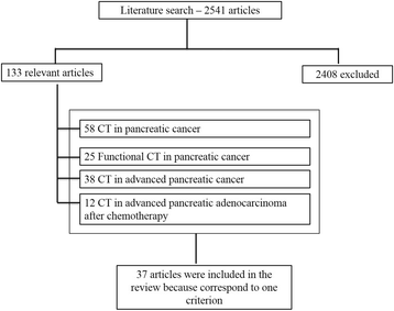
Fig. 1
Fig. 1
Included and excluded studies in systematic review
Fig. 2
Fig. 2
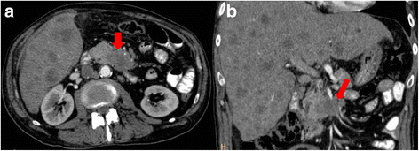
CT scan in axial plane (a) and coronal plane, Multiplanar Reconstruction-MPR (b), during pancreatic phase of dynamic contrast study. Body-tail adenocarcinoma (arrow)
Fig. 3
Fig. 3
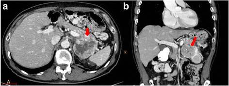
CT scan in axial plane (a) and coronal plane, MPR (b), during portal phase of dynamic contrast study. Tail pancreatic adenocarcinoma that infiltrates vascular hilum of the spleen
Fig. 4
Fig. 4
Maximum Intensity Projection (MIP); arrow shows vascular infiltration
Fig. 5
Fig. 5
CT scan in axial plane during pancreatic phase of dynamic contrast study. Isodense pancreatic adenocarcinoma (arrow)
Fig. 6
Fig. 6
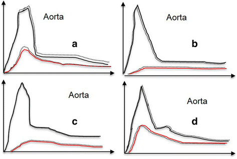
a Type A curve wash-in followed by wash-out (normal parenchyma); b type B curve-low wash-in, followed by plateau or increasing density, without wash-out (adenocarcinoma); c type C curve-low wash-in, followed by at least a slight wash-out (chronic pancreatitis); d type D curve-brisk wash-in, followed by clear wash-out (endocrine tumor)

References

    1. Siegel R, Naishadham D, Jemal A. Cancer statistics, 2013. CA Cancer J Clin. 2013;63:11–30. doi: 10.3322/caac.21166. - DOI - PubMed
    1. Granata V, Fusco R, Piccirillo M, Palaia R, Lastoria S, Petrillo A, Izzo F. Feasibility and safety of intraoperative electrochemotherapy in locally advanced pancreatic tumor: a preliminary experience. Eur J Inflamm. 2014;12(3):467–477.
    1. Lee ES, Lee JM. Imaging diagnosis of pancreatic cancer: a state-of-the-art review. World J Gastroenterol. 2014;20(24):7864–7877. doi: 10.3748/wjg.v20.i24.7864. - DOI - PMC - PubMed
    1. Granata V, Fusco R, Piccirillo M, Palaia R, Petrillo A, Lastoria S, Izzo F. Electrochemotherapy in locally advanced pancreatic cancer: preliminary results. Int J Surg. 2015;18:230–236. doi: 10.1016/j.ijsu.2015.04.055. - DOI - PubMed
    1. Chang J, Schomer D, Dragovich T. Anatomical, physiological, and molecular imaging for pancreatic cancer: current clinical use and future implications. Biomed Res Int. 2015;2015:269641. - PMC - PubMed

LinkOut - more resources