

doi: 10.1155/2016/1490181.
Epub 2016 Nov 7.
MELAS Syndrome with Cardiac Involvement: A Multimodality Imaging Approach
Affiliations
- PMID: 27891257
- PMCID: PMC5116498
- DOI: 10.1155/2016/1490181
Item in Clipboard
MELAS Syndrome with Cardiac Involvement: A Multimodality Imaging Approach
Case Rep Cardiol.
2016.
Abstract
A 49-year-old man presented with chest pain, dyspnea, and lactic acidosis. Left ventricular hypertrophy and myocardial fibrosis were detected. The sequencing of mitochondrial genome (mtDNA) revealed the presence of A to G mtDNA point mutation at position 3243 (m.3243A>G) in tRNALeu(UUR) gene. Diagnosis of cardiac involvement in a patient with Mitochondrial Encephalomyopathy, Lactic Acidosis, and Stroke-like episodes syndrome (MELAS) was made. Due to increased risk of sudden cardiac death, cardioverter defibrillator was implanted.
Conflict of interest statement
The authors report no financial relationships or competing interests regarding the content herein.
Figures
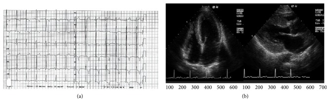
(a) Electrocardiogram showing short PR interval and left ventricular (LV) hypertrophy. (b) Echocardiogram showing concentric, nonobstructive LV hypertrophy.
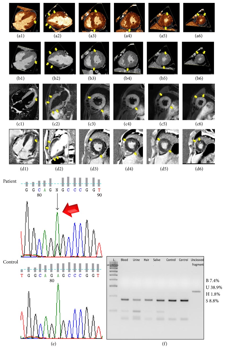
Color-coded (a1–a6) and merged gray-scale (b1–b6) late-enhancement Dual-Energy CT perfusion maps in four-chamber (a1, b1), two-chamber (a2, b2), and short-axis views from base to apex (a3–a6 and b3–b6, resp.) showing the left ventricular (LV) hypertrophy and diffuse, patchy, nonischemic (predominantly intramural), late-enhancement (arrows). T2-STIR MRI imaging (c1–c6) and phase sensitive T1-weighted inversion recovery late-enhancement MRI images (d1–d6) in four-chamber (c1, d1), two-chamber (c2, d2), and short-axis views from base to apex (c3–c6 and d3–d6, resp.) demonstrated diffuse, patchy, nonischemic (predominantly intramural) myocardial edema and late-enhancement consistent with necrosis/fibrosis with a high level of concordance with Dual-Energy CT. (e) Sequence chromatograph of the tRNA
Leu(UUR) region flanking the m.3243A>G mutation (arrow) in blood sample from the proband and in a wild type sample (Ctr). (f) PCR-Restriction Fragment Length Polymorphism analysis showed the variable mutant load in patient's peripheral tissues. B: blood; U: urine; H: hair; S: saliva.
References
LinkOut - more resources
Full Text Sources
Other Literature Sources
