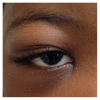
Orbital Infarction due to Sickle Cell Disease without Orbital Pain
- PMID: 27891273
- PMCID: PMC5116510
- DOI: 10.1155/2016/5867850
Orbital Infarction due to Sickle Cell Disease without Orbital Pain
Abstract
Sickle cell disease is a hemoglobinopathy that results in paroxysmal arteriolar occlusion and tissue infarction that can manifest in a plurality of tissues. Rarely, these infarcted crises manifest in the bony orbit. Orbital infarction usually presents with acute onset of periorbital tenderness, swelling, erythema, and pain. Soft tissue swelling can result in proptosis and attenuation of extraocular movements. Expedient diagnosis of sickle cell orbital infarction is crucial because this is a potentially sight-threatening entity. Diagnosis can be delayed since the presentation has physical and radiographic findings mimicking various infectious and traumatic processes. We describe a patient who presented with sickle cell orbital crisis without pain. This case highlights the importance of maintaining a high index of suspicion in patients with known sickle cell disease or of African descent born outside the United States in a region where screening for hemoglobinopathy is not routine, even when the presentation is not classic.
Conflict of interest statement
The authors declare that there are no competing interests regarding the publication of this paper.
Figures


References
-
- Gill F. M., Sleeper L. A., Weiner S. J., et al. Clinical events in the first decade in a cohort of infants with sickle cell disease. Cooperative Study of Sickle Cell Disease. Blood. 1995;86(2):776–783. - PubMed
-
- Serjeant G. R. Sickle Cell Disease. 2nd. Oxford, UK: Oxford University Press; 1992.
Grants and funding
LinkOut - more resources
Full Text Sources
Other Literature Sources

