. 2016:2016:5946520.
doi: 10.1155/2016/5946520. Epub 2016 Nov 7.

Size and Proportions of Slow-Twitch and Fast-Twitch Muscle Fibers in Human Costal Diaphragm

Affiliations

Size and Proportions of Slow-Twitch and Fast-Twitch Muscle Fibers in Human Costal Diaphragm

Marija Meznaric et al. Biomed Res Int. 2016.

Abstract

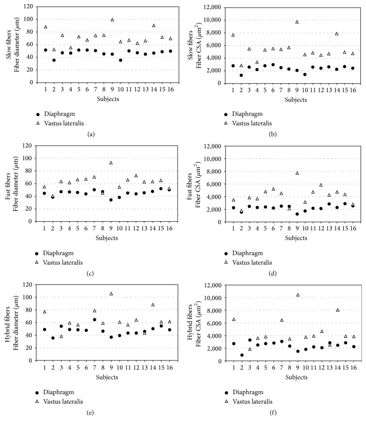
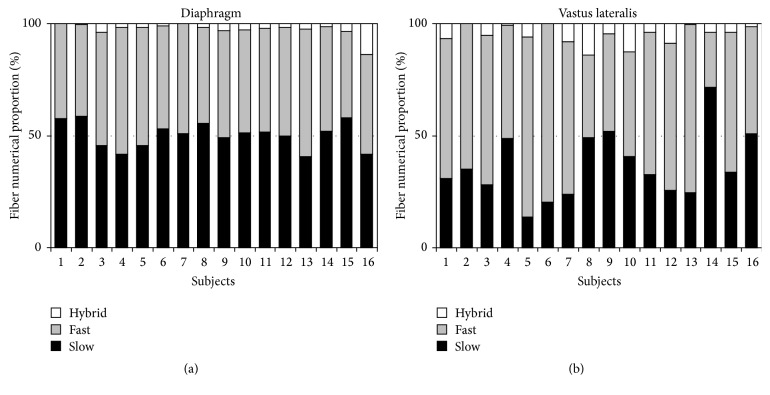
Smaller diaphragmatic motor unit potentials (MUPs) compared to MUPs of limb muscles lead to the hypothesis that diaphragmatic muscle fibers, being the generators of MUPs, might be also smaller. We compared autopsy samples of costal diaphragm and vastus lateralis of healthy men with respect to fibers' size and expression of slow myosin heavy chain isoform (MyHC-1) and fast 2A isoform (MyHC-2A). Diaphragmatic fibers were smaller than fibers in vastus lateralis with regard to the mean minimal fiber diameter of slow-twitch (46.8 versus 72.2 μm, p < 0.001), fast-twitch (45.1 versus 62.4 μm, p < 0.001), and hybrid fibers (47.3 versus 65.0 μm, p < 0.01) as well as to the mean fiber cross-sectional areas of slow-twitch (2376.0 versus 5455.9 μm2, p < 0.001), fast-twitch (2258.7 versus 4189.7 μm2, p < 0.001), and hybrid fibers (2404.4 versus 4776.3 μm2, p < 0.01). The numerical proportion of slow-twitch fibers was higher (50.2 versus 36.3%, p < 0.01) in costal diaphragm and the numerical proportion of fast-twitch fibers (47.2 versus 58.7%, p < 0.01) was lower. The numerical proportion of hybrid fibers did not differ. Muscle fibers of costal diaphragm have specific characteristics which support increased resistance of diaphragm to fatigue.


Conflict of interest statement

The authors declare that there are no competing interests regarding the publication of this paper.

Figures

Figure 1
Figure 1
Phenotyping of muscle fibers by the expression of slow and fast MyHCs in costal diaphragm and vastus lateralis muscle. Fibers labeled by dots are slow-twitch fibers, fibers labeled by open circles are fast-twitch fibers, and hybrid fibers are labeled by arrows. Muscle fibers not stained by antibodies to slow MyHC are stained heavily or intermediately by antibodies to fast MyHC. Bar = 50 μm.
Figure 2
Figure 2
Variability between subjects with regard to fiber diameters and fiber cross-sectional areas. CSA = cross-sectional area. Slow-twitch fibers (a, b) of costal diaphragm were smaller with regard to diameters and fiber cross-sectional areas than those of vastus lateralis muscle in all subjects. Fast-twitch fibers of costal diaphragm (c, d) were in 13/16 subjects smaller and in 3/16 subjects of similar size compared to those of vastus lateralis. Hybrid fibers (e, f) of costal diaphragm were in 12/16 subjects smaller, in 1/16 of similar size, and in 1/16 bigger than those of vastus lateralis. In 2/16 comparison was not possible, since hybrid fibers were absent in vastus lateralis muscle.
Figure 3
Figure 3
Variability between subjects with regard to the numerical proportions of fiber types. Numerical proportion of slow-twitch fibers of costal diaphragm (a) was in all subjects around 50%, while in vastus lateralis muscle (b) this was the case only in 4/16 subjects and in the majority (11/16) slow-twitch fibers constitute about one-third of muscle fibers of vastus lateralis. In 1/16 subjects the proportion of slow-twitch fibers was around 70%. The proportion of hybrid fibers was small but varied considerably in both muscles; in costal diaphragm minimum was 0.1% and maximum 13.4% and in vastus lateralis minimum was 0.0% and maximum 14%.

References

    1. Podnar S., Resman-Gašperšič A. Quantitative motor unit potential analysis in the diaphragm: a normative study. Muscle & Nerve. 2008;37(4):518–521. doi: 10.1002/mus.20939. - DOI - PubMed
    1. Nandedkar S., Stalberg E., Sanders D. Quantitative EMG. In: Dumitru D., Amato A. A., Zwarts M., editors. Electrodiagnostic Medicine. 2nd. Philadelphia, Pa, USA: Hanley & Belfus; 2002. pp. 293–356.
    1. Sauleda J., Gea J., Orozco-Levi M., et al. Structure and function relationships of the respiratory muscles. European Respiratory Journal. 1998;11(4):906–911. doi: 10.1183/09031936.98.11040906. - DOI - PubMed
    1. Mizuno M. Human respiratory muscles: fibre morphology and capillary supply. The European Respiratory Journal. 1991;4(5):587–601. - PubMed
    1. Polla B., D'Antona G., Bottinelli R., Reggiani C. Respiratory muscle fibres: specialisation and plasticity. Thorax. 2004;59(9):808–817. doi: 10.1136/thx.2003.009894. - DOI - PMC - PubMed

LinkOut - more resources