. 2017 May;26(5):423-430.
doi: 10.1111/exd.13256. Epub 2017 Feb 20.

SVEP1 plays a crucial role in epidermal differentiation

Affiliations

SVEP1 plays a crucial role in epidermal differentiation

Liat Samuelov et al. Exp Dermatol. 2017 May.

Abstract

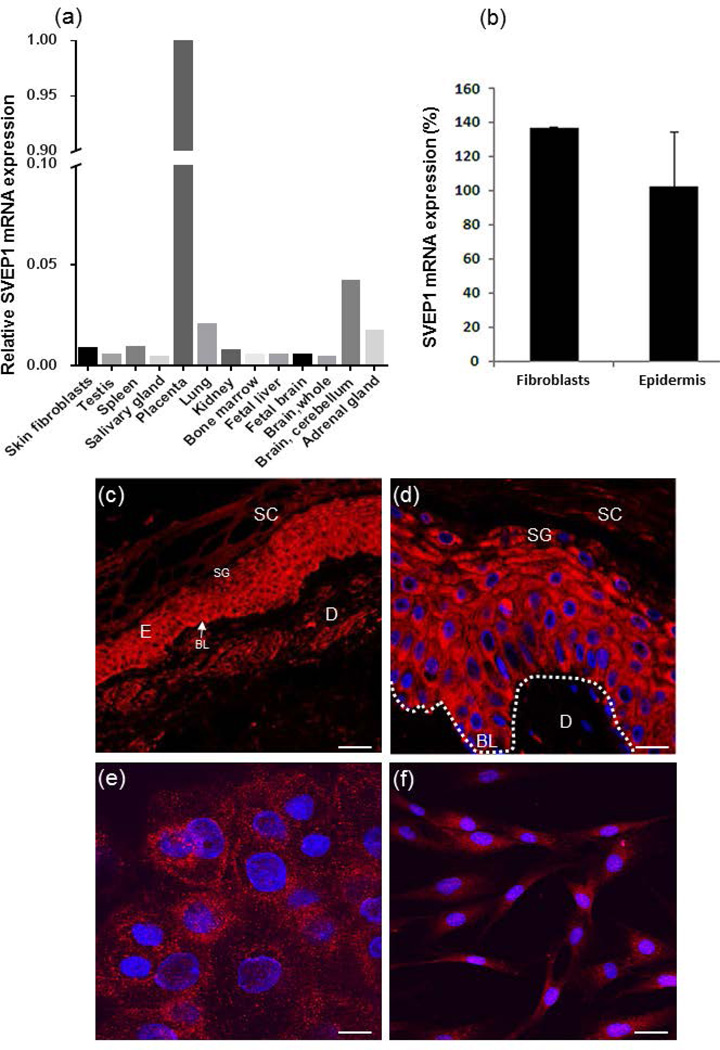
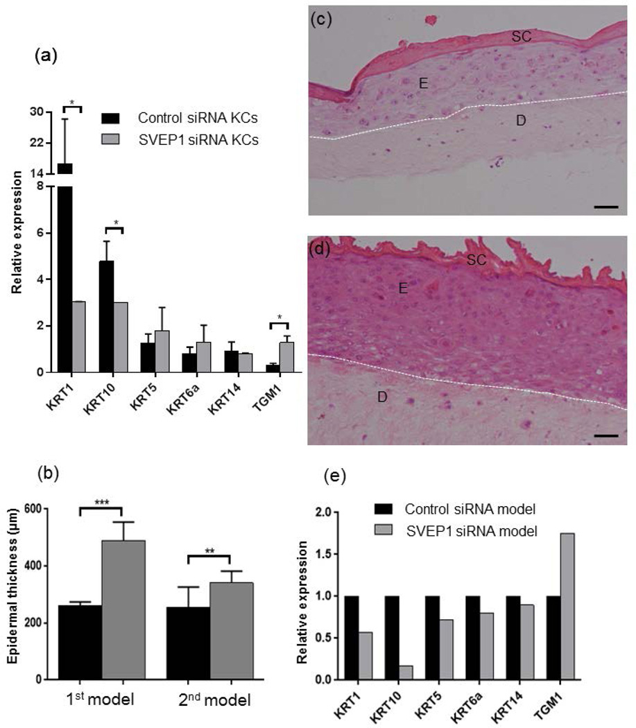
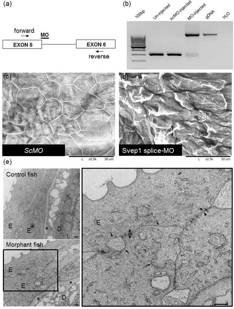
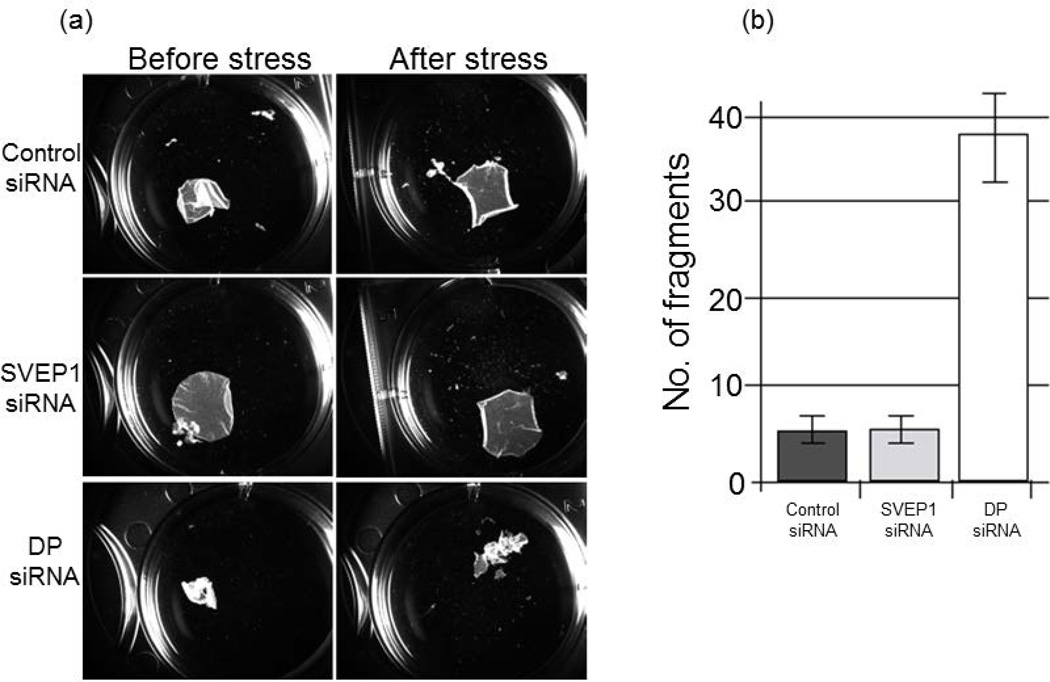
SVEP1 is a recently identified multidomain cell adhesion protein, homologous to the mouse polydom protein, which has been shown to mediate cell-cell adhesion in an integrin-dependent manner in osteogenic cells. In this study, we characterized SVEP1 function in the epidermis. SVEP1 was found by qRT-PCR to be ubiquitously expressed in human tissues, including the skin. Confocal microscopy revealed that SVEP1 is normally mostly expressed in the cytoplasm of basal and suprabasal epidermal cells. Downregulation of SVEP1 expression in primary keratinocytes resulted in decreased expression of major epidermal differentiation markers. Similarly, SVEP1 downregulation was associated with disturbed differentiation and marked epidermal acanthosis in three-dimensional skin equivalents. In contrast, the dispase assay failed to demonstrate significant differences in adhesion between keratinocytes expressing normal vs low levels of SVEP1. Homozygous Svep1 knockout mice were embryonic lethal. Thus, to assess the importance of SVEP1 for normal skin homoeostasis in vivo, we downregulated SVEP1 in zebrafish embryos with a Svep1-specific splice morpholino. Scanning electron microscopy revealed a rugged epidermis with perturbed microridge formation in the centre of the keratinocytes of morphant larvae. Transmission electron microscopy analysis demonstrated abnormal epidermal cell-cell adhesion with disadhesion between cells in Svep1-deficient morphant larvae compared to controls. In summary, our results indicate that SVEP1 plays a critical role during epidermal differentiation.

Keywords: SVEP1; epidermal differentiation; integrin α9β1; zebrafish.


Conflict of interest statement

Conflicts of interests

The authors declare no conflicts of interests.

Figures

Figure 1
Figure 1. SVEP1 expression in human tissues
(a) SVEP1 mRNA expression was evaluated using qRT-PCR in normal human tissues. Results are expressed as relative to gene expression in the placenta normalized to ACTB RNA levels; (b) SVEP1 mRNA expression in cultured human KCs and fibroblasts. Results are expressed as % of expression relative to keratinocytes and are normalized to ACTB RNA levels; (c) SVEP1 immunostaining of skin biopsies obtained from healthy individual reveals SVEP1 expression in all epidermal layers and in dermal fibroblasts residing in the upper dermis; (d) A higher magnification demonstrates increased expression of SVEP1 in the basal and suprabasal layers of the epidermis (bars; c = 100µm, d = 40µm). Dotted lines represent epidermal-dermal junction and localization of the basement membrane; E-epidermis; D-dermis; SC-stratum corneum; SG-stratum granulosum; BL-basal layer; (e–f) KCs (e) and fibroblasts (f) isolated from skin biopsies obtained from healthy individuals demonstrate SVEP1 expression most prominent in the peripheral cytoplasmic and nuclear compartments of the cells (bar = 40 µm).
Figure 2
Figure 2. SVEP1 knockdown in primary KCs and organotypic cell cultures
(a) Gene expression was assessed by qRT-PCR in SVEP1-silenced primary KCs (KCs) compared to control small interfering RNA (siRNA) treated KCs. Following siRNA transfection, KCs were cultured with and without calcium (1.5mM) for 96 hours. Results are expressed as relative gene expression in KCs cultured at high vs. low calcium concentration (t-test; *p<0.05). Results were normalized to GAPDH RNA levels; (b–e) Human primary KCs and fibroblasts transfected with SVEP1 siRNA or control siRNA were used to generate skin equivalents. The experiment was repeated twice under identical conditions (the results of the first experiment are designated as “1st model” while the results of the second experiment are designated as “2nd model”). Punch biopsies were obtained from skin equivalents at day 10 and stained for hematoxylin and eosin (H&E; bar = 50µm). Epidermal thickness was evaluated in SVEP1-deficient and control siRNA treated skin equivalents, in both experiments (b). Note significant acanthosis in skin equivalents downregulated for SVEP1 (d) compared to control (c). Dotted lines represent epidermal-dermal junction. E-epidermis; D-dermis; SC-stratum corneum; (e)RNA was extracted from punch biopsies derived from skin equivalents and gene expression was assessed using qRT-PCR. Results are expressed gene expression in down-regulated skin equivalents relative to control skin equivalents. Data were normalized to GAPDH RNA levels (t-test; *p<0.05, **p<0.01.
Figure 3
Figure 3. Svep1 knockdown in a zebrafish model
(a–b) Knockdown of Svep1 by a splice site morphilino (MO). The morpholino targets the splice donor site at the exon 5/intron 5 border of zebrafish Svep1 gene (a). The consequences of MO on Svep1 pre-mRNA processing was determined by RT-PCR using primers located in exons 5 and 6 (b). scMO-standard control morpholino; gDNA-genomic DNA; (c–d) Scanning electron microscopy analysis of the skin of the tail of a control larvae injected with the global standard control morpholino (scMO) shows the presence of KCs with well-demarcated cell-cell borders and containing microridges (c) while the morphant larvae injected with a splice site morpholino for Svep1 gene reveals rugged epidermis with perturbed microridge formation in the center of the KCs (d); (e) Transmission electron microscopy demonstrates abnormal epidermal cell-cell adhesion with disadhesion between the cells in the Svep1 morphant larvae compared to control. Arrows mark the separation of cell-cell contacts while asterisks mark the basement membrane. Arrowheads point to the presence of normal appearing desmosomes. (bar= 500nm). E-epidermis; D-dermis.
Figure 4
Figure 4. Intact intercellular adhesion in epithelial sheets in the absence of SVEP1
(a) Electroporation of normal human foreskin-derived keratinocytes was performed using siRNA targeting SVEP1, DSP encoding desmoplakin (DP) or a scrambled control siRNA. DP is used as a positive control for decreased intercellular adhesion. 72 h following siRNA treatment, cell monolayers were seeded in triplicate into 6-well plates. 24 h after reaching confluency, cultures were washed twice in DPBS and then incubated in 2 ml of dispase (2.4 U/ml) for 30 min; (b) Released monolayers were subjected to mechanical stress to induce fragmentation. Quantification of the number of total particles in each well between each condition is shown as an average from the triplicate wells after stress. Fragments were counted using a dissecting microscope (MZ6; Leica). Results are representative of three experiments each performed in triplicate.

References

    1. Sumigray KD, Lechler T. Cell adhesion in epidermal development and barrier formation. Current topics in developmental biology. 2015;112:383–414. - PMC - PubMed
    1. Simpson CL, Patel DM, Green KJ. Deconstructing the skin: cytoarchitectural determinants of epidermal morphogenesis. Nature reviews Molecular cell biology. 2011;12:565–580. - PMC - PubMed
    1. Samuelov L, Sprecher E. Inherited desmosomal disorders. Cell and tissue research. 2015;360:457–475. - PubMed
    1. Glait-Santar C, Pasmanik-Chor M, Benayahu D. Expression pattern of SVEP1 alternatively-spliced forms. Gene. 2012;505:137–145. - PubMed
    1. Shur I, Socher R, Hameiri M, et al. Molecular and cellular characterization of SEL-OB/SVEP1 in osteogenic cells in vivo and in vitro. Journal of cellular physiology. 2006;206:420–427. - PubMed

Publication types

Substances