. 2017 Jan;23(1):39-48.
doi: 10.1038/nm.4240. Epub 2016 Nov 28.

Thalamic miR-338-3p mediates auditory thalamocortical disruption and its late onset in models of 22q11.2 microdeletion

Affiliations

Thalamic miR-338-3p mediates auditory thalamocortical disruption and its late onset in models of 22q11.2 microdeletion

Sungkun Chun et al. Nat Med. 2017 Jan.

Abstract

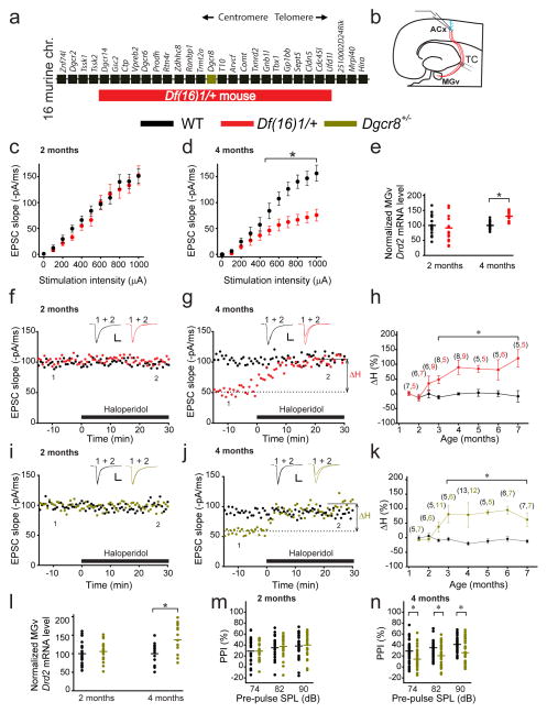
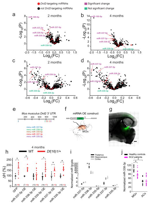
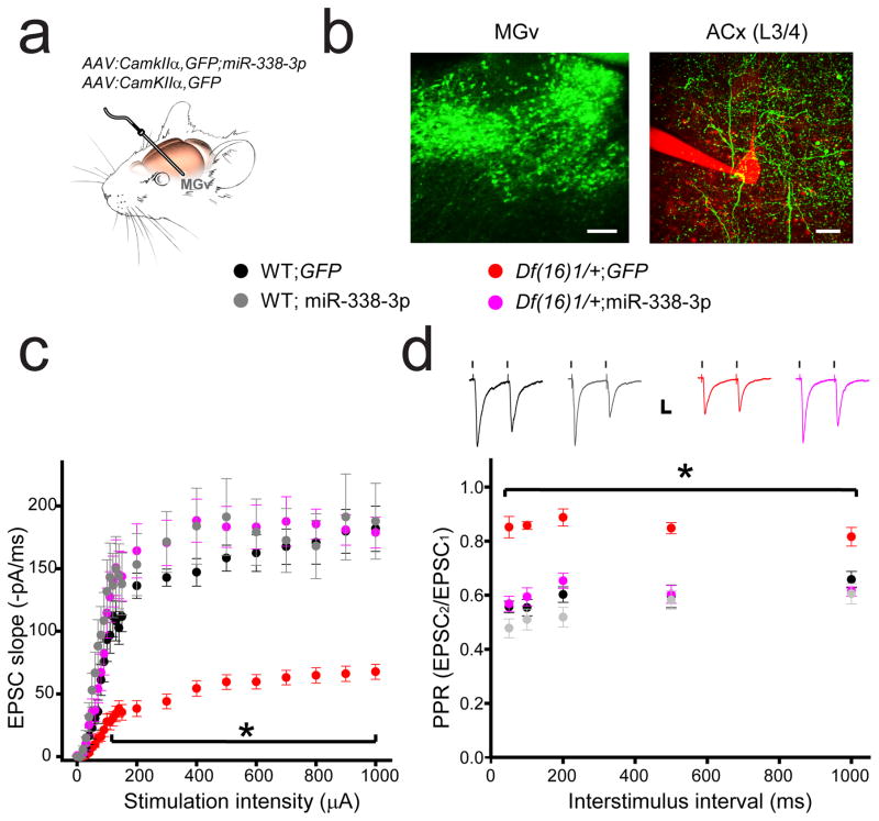
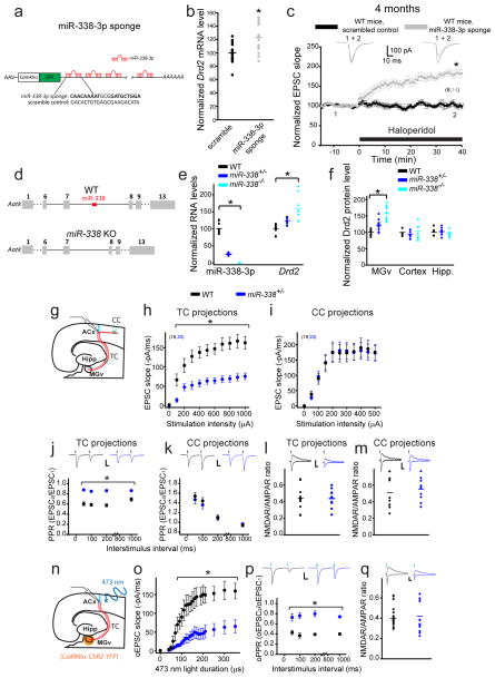
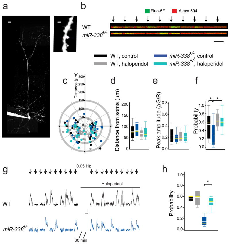
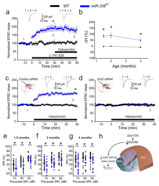
Although 22q11.2 deletion syndrome (22q11DS) is associated with early-life behavioral abnormalities, affected individuals are also at high risk for the development of schizophrenia symptoms, including psychosis, later in life. Auditory thalamocortical (TC) projections recently emerged as a neural circuit that is specifically disrupted in mouse models of 22q11DS (hereafter referred to as 22q11DS mice), in which haploinsufficiency of the microRNA (miRNA)-processing-factor-encoding gene Dgcr8 results in the elevation of the dopamine receptor Drd2 in the auditory thalamus, an abnormal sensitivity of thalamocortical projections to antipsychotics, and an abnormal acoustic-startle response. Here we show that these auditory TC phenotypes have a delayed onset in 22q11DS mice and are associated with an age-dependent reduction of miR-338-3p, a miRNA that targets Drd2 and is enriched in the thalamus of both humans and mice. Replenishing depleted miR-338-3p in mature 22q11DS mice rescued the TC abnormalities, and deletion of Mir338 (which encodes miR-338-3p) or reduction of miR-338-3p expression mimicked the TC and behavioral deficits and eliminated the age dependence of these deficits. Therefore, miR-338-3p depletion is necessary and sufficient to disrupt auditory TC signaling in 22q11DS mice, and it may mediate the pathogenic mechanism of 22q11DS-related psychosis and control its late onset.


Conflict of interest statement

The authors declare no conflict of interest.

Figures

Fig. 1
Fig. 1. Adult onset of antipsychotics sensitivity and synaptic transmission disruption in auditory TC projections of mouse models of 22q11DS
(a) Map of 22q11DS orthologs deleted in Df(16)1/+ mice. (b) Illustration of voltage-clamp recordings of thalamorecipient L3/4 pyramidal neurons in TC slices. TC projections are shown in red. ACx, auditory cortex; TC, thalamocortical; MGv, ventral part of the medial geniculate nuclei. (c, d) Input–output relations between stimulation intensity and EPSCs at TC projections in the ACx of 2- (c, F(1,37)=0.967, p=0.338) or 4-month-old (d, F(1,46)=11.56, *p<0.001) WT (20 and 23 neurons, respectively) and Df(16)1/+ mice (19 and 25 neurons, respectively). (e) Drd2 transcript levels in the MGV of 2- and 4-month-old WT and Df(16)1/+ mice (2 months: 6 mice of each genotype, measured in triplicates, U=126; p=0.261; 4 months: 5 mice of each genotype, measured in triplicates, t(28)= –5.78; *p <0.001). (f, g) The effect of haloperidol on TC EPSCs in 2- (f) and 4-month-old (g) WT and Df(16)1/+ littermates. Haloperidol-induced percentage change (ΔH) in the slope of TC EPSCs relative to baseline (before haloperidol application; dashed line). (h) The ΔH as a function of mouse age in WT and Df(16)1/+ littermates. (i, j) The effect of haloperidol on TC EPSCs in 2- (i) and 4-month-old (j) WT and Dgcr8+/− littermates. (f, g, i, j) Scale bars, 50 pA, 10 ms. Insets show representative EPSCs before (1) and after (2) haloperidol application. (k) The ΔH as a function of mouse age in WT and Dgcr8+/− littermates. (h, k) The number of cells recorded at each age is shown in parentheses above the plots. *p <0.01 (two-tailed t-test or Mann-Whitney Rank Sum U test). (l) Average Drd2 mRNA levels normalized to Gapdh in the auditory thalamus of 2- and 4-month-old WT and Dgcr8+/− littermates (2 months: 7 mice of each genotype; 4 months: 4 WT mice, 5 Dgcr8+/− mice, measured in triplicates).The t(24)=–2.44; *p=0.022, two-tailed t-test. (m, n) Mean PPI of maximal acoustic-startle response in 2- (m) and 4-month-old (n) WT and Dgcr8+/− littermates (2 months: 23 WT mice and 22 Dgcr8+/− mice; 4 months: 36 WT mice and 41 Dgcr8+/− mice). *p <0.05 (two-tailed t-test or Mann-Whitney Rank Sum U test). SPL (sound pressure level). (c, d, h, k, m, n) Data are represented as the mean ± SEM. (e, l–n) Horizontal lines represent the mean values.
Fig. 2
Fig. 2. Identification of Drd2-targeting miR-338-3p in the auditory thalamus
(a–d) Volcano plots of miRNA microarray data from the auditory thalamus of 2- (a, c) and 4-month-old (b, d) WT and Df(16)1/+ (a, b) and WT and Dgcr8+/− (c, d) male littermates. The difference between miRNA levels in WT and mutants was considered significant if p <0.01. Symbol size represents the miRNA expression level in the microarray. Note, miR-338-3p had the highest expression among all predicted Drd2-targeting miRNAs. (e) Diagram of the mouse Drd2 3′UTR (XM_006509996.2) with seed sites for the 5 miRNAs indicated. (f) Experimental design of a recombinant AAV encoding a chimeric construct overexpressing an miRNA of interest (top) injected into the mouse MGv (bottom). (g) GFP expressed specifically in the auditory TC projections after in vivo injection of recombinant AAV. Scale bar, 500 μm. (h) Haloperidol sensitivity of TC projections in 4-month-old WT and Df(16)1/+ mice injected with AAVs encoding different miRNAs or GFP. The number of cells recorded is shown in parentheses. *p <0.01 (two-tailed t-test or Mann-Whitney Rank Sum U test). (i) Relative average levels of miRNA expression in the thalamus, hippocampus, and cortex of WT mice (5 mice, run in triplicates). Data normalized to the average of three housekeeping genes: U6, snoRNA202, and snoRNA234. Only miR-338-3p shows enrichment in the thalamus. H(2)=18.85, *p <0.001. (j) Mean relative miR-338-3p levels (normalized to U6) in the postmortem MGv and ACx tissues from healthy controls and patients with schizophrenia (SCZ) (MGv: 7 controls and 7 patients (t(40)=4.56; *p <0.001); ACx: 8 controls and 8 patients (U=278; p=0.845), measured in triplicates). *p <0.05.
Fig. 3
Fig. 3. Replenishment of miR-338-3p in the auditory thalamus rescues deficits in synaptic transmission and presynaptic neurotransmitter release at TC projections of 22q11DS mouse models
(a) In vivo infection of MGv relay neurons with AAV-GFP-miR-338-3p or AAV-GFP. (b) GFP expression (green) in cell bodies in the MGv (left; scale bar, 100 μm) and in projections to the thalamorecipient L3/4 layer of the ACx (right; scale bar, 20 μm). A patch pipette and part of an L3/4 pyramidal neuron filled with Alexa 594 are shown in red. (c, d) Input–output relations between stimulation intensity and EPSCs (c) and PPR (d) at TC projections in the ACx of 4- to 5-month-old WT and Df(16)1/+ mice injected with either AAV-GFP-miR-338-3p or AAV-GFP. Insets show representative EPSCs. Scale bar, 20 ms, 50 pA. (c) 16 WT;GFP neurons, 10 WT; miR-338-3p neurons, 20 Df(16)1/+;GFP neurons, 13 Df(16)1/+; miR-338-3p neurons. F(3,24)=268.7, *p<0.001. (d) 17 WT;GFP neurons, 12 WT; miR-338-3p neurons, 19 Df(16)1/+;GFP neurons, 14 Df(16)1/+; miR-338-3p neurons. F(3,4)=107.2, *p<0.001. Data are represented as the mean ± SEM.
Fig. 4
Fig. 4. The depletion or knockout of miR-338 replicates the TC deficiency of Df(16)1/+ mice
(a) AAV expressing an miR-338-3p sponge construct with multiple binding sites to miR-338-3p in the GFP 3′UTR, under control of the CamKIIα promoter. Sequences for the miR-338-3p sponge and the scrambled control are shown below. Bold text indicates seed-site sequence. (b) Relative Drd2 mRNA levels after infection of MGv excitatory neurons in WT mice with an AAV encoding a scrambled control (5 mice, run in triplicates) or miR-338-3p sponge (6 mice, run in triplicates), U=53, *p=0.005. (c) Normalized mean TC EPSCs before and after application of haloperidol in WT mice after infection of MGv neurons with AAVs encoding a scrambled control (6 neurons) or miR-338-3p sponge (14 neurons). Insets show representative EPSCs. *p <0.001 (two-tailed t-test). (d) Generation of miR-338 knockout (KO) mice. (e) Normalized levels of miR-338-3p and Drd2 in the auditory thalamus of WT (miR-338-3p: 3 mice, Drd2: 5 mice), miR-338+/− (miR-338-3p: 4 mice, Drd2: 6 mice) and miR-338−/− mice (miR-338-3p: 4 mice, Drd2: 4 mice). Run in triplicates. miR-338-3p: H(2)=26.5, *p <0.001; Drd2: H(2)=25.2, *p <0.001. (f) Normalized levels of Drd2 protein in the auditory thalamus (H(2)=23.3, *p<0.01), cortex (H(2)=1.21, p=0.544), and hippocampus (Hipp.) (t(2)=0.4, p=0.674) of WT (3 mice), miR-338+/− (4 mice), and miR-338−/− mice (4 mice). Run in duplicates or triplicates. (g) Simultaneous recordings of EPSCs in L3/4 pyramidal neurons evoked by electrical stimulation of the thalamocortical (TC) and corticocortical (CC) projections. (h, i) Input–output relations between electrical stimulation intensity and EPSCs at TC projections (h, F(1,37)=26.9, *p <0.001) and CC projections (i, F(1,37)=0.002, p=0.964) in the ACx of 4-month-old WT mice (19 neurons) and miR-338+/− mice (20 neurons). PPR ratio (j, k) and NMDAR/AMPAR ratio (l, m) of electrically evoked EPSCs measured at TC projections (j, l) and CC projections (k, m) of 4-month-old WT (26, 23, 10, 9 neurons, respectively) and miR-338+/− mice (22, 19, 12, 10 neurons, respectively). (j) *p <0.001 (two-tailed t-test); (k) p >0.05 (two-tailed t-test); (l) t(20)=0.03, p=0.974; (m) t(17)=–0.576, p=0.572. (n) Optogenetic experiments in TC slices. ChR2 was expressed in the MGv, under control of the CamKIIα promoter. (o–q) Input–output relations (o), PPR (p), and NMDAR/AMPAR ratio (q) of optically evoked EPSCs (oEPSC) measured at TC projections of 4-month-old WT (10, 16, 14 neurons, respectively) and miR-338+/− mice (9, 16, 17 neurons, respectively). (o) F(1,17)=11.25, *p=0.004; (p) *p <0.001 (two-tailed t-test); (q) U=98, p=0.296. Insets show representative AMPAR-mediated (–70 mV holding membrane potential) and NMDAR-mediated (+40 mV holding membrane potential) EPSC and oEPSC traces. Scale bars, 20 ms, 50 pA. (c, h–k, o, p) Data are represented as the mean ± SEM. (b, e, f, l, m, q) Horizontal lines represents the mean values. *p <0.01.
Fig. 5
Fig. 5. Probability of glutamate release is reduced at TC projections of miR-338+/− mice
(a) An L3/4 pyramidal neuron filled with Fluo-5F and Alexa 594 through a patch pipette (left; scale bar, 10 μm) to visualize synaptically evoked calcium transients inside dendritic spines (right; scale bar, 1μm). Yellow line represents the line scan. (b) Calcium transients in a dendritic spine in response to a single thalamic stimulation (arrows) repeated 10 times at 0.05–0.1 Hz. Scale bar, 0.1 ΔG/R, 200 ms. (c) Location of active TC inputs on dendritic trees of L3/4 pyramidal neurons spines. (0;0), soma coordinates (apical dendrites pointing upwards). (d–f) Average distances from the soma to active TC inputs (d), calcium transient peak amplitudes (e), and probabilities (f) in response to 10 to 20 single TC stimulations in WT and miR-338+/− slices not treated (WT: 27 spines; miR-338+/−: 32 spines) or treated (WT: 32 spines; miR-338+/−: 41spines) with haloperidol. (d) F(3)=1.05, p=0.373; (e) H(3)=0.644, p=0.886; (f) H(3)=31.51, *p <0.001. (g, h) Representative traces (g) and average probability (h) of calcium transients in the same dendritic spines before and after haloperidol application in WT (5 spines) and miR-338+/− mice (9 spines). H(3)=14.5, *p=0.002. (d–f, h) Data are represented as the mean (white lines), median (yellow lines), 10th, 25th, 75th, and 90th percentiles.
Fig. 6
Fig. 6. Deletion of miR-338 in mice eliminates age dependency for antipsychotics sensitivity and replicates 22q11DS phenotypes
(a) Average TC EPSCs before (1) and during (2–3) application of the Drd2-specific inhibitor L-741,626 and haloperidol in 4-month-old WT (6 neurons) and miR-338+/− mice (9 neurons). (b) Mean haloperidol sensitivity (ΔH) in WT (10, 7, 6 neurons at 1.5, 2, and 4 months, respectively) and miR-338+/− mice (12 neurons at each age) between 1.5 and 4 months of age. *p <0.001 (two-tailed t-test). (c, d) Mean TC EPSCs before (1) and after (2) haloperidol in 2-month-old WT (10 control siRNA neurons and 9 Drd2 siRNA neurons) and miR-338+/− mice (14 control siRNA neurons and 10 Drd2 siRNA neurons) that received control (c) or Drd2 siRNA injected into their MGv (d). Insets show representative EPSCs. (e–g) Mean PPI of maximal acoustic startle response in 1.5- (e), 2- (f), and 4-month-old (g) WT (22, 22, 21 mice, respectively) and miR-338+/− littermates (21, 21, 20 mice, respectively). (e) F(5)=21.648, *p<0.001; (f) H(5)=39.887, *p<0.001; (g) H(5)=17.348, *p=0.004. SPL, sound pressure level. (h) Model of TC disruption in 22q11DS. DGCR8-dependent depletion of the thalamus-enriched miR-338-3p leads to an increase in DRD2 level in the auditory thalamus (MGv) and disruption of thalamocortical synaptic transmission to the auditory cortex (ACx) later in life.

References

    1. Dierks T, et al. Activation of Heschl’s gyrus during auditory hallucinations. Neuron. 1999;22:615–621. - PubMed
    1. Silbersweig DA, et al. A functional neuroanatomy of hallucinations in schizophrenia. Nature. 1995;378:176–179. - PubMed
    1. Horga G, Schatz KC, Abi-Dargham A, Peterson BS. Deficits in predictive coding underlie hallucinations in schizophrenia. J Neurosci. 2014;34:8072–8082. - PMC - PubMed
    1. Javitt DC, Sweet RA. Auditory dysfunction in schizophrenia: integrating clinical and basic features. Nat Rev Neurosci. 2015;16:535–550. - PMC - PubMed
    1. Chun S, et al. Specific disruption of thalamic inputs to the auditory cortex in schizophrenia models. Science. 2014;344:1178–1182. - PMC - PubMed

Publication types

MeSH terms