. 2016 Nov 1;57(14):6407-6415.
doi: 10.1167/iovs.16-20177.

Potential of Small Molecule-Mediated Reprogramming of Rod Photoreceptors to Treat Retinitis Pigmentosa

Affiliations

Potential of Small Molecule-Mediated Reprogramming of Rod Photoreceptors to Treat Retinitis Pigmentosa

Paul A Nakamura et al. Invest Ophthalmol Vis Sci. .

Abstract

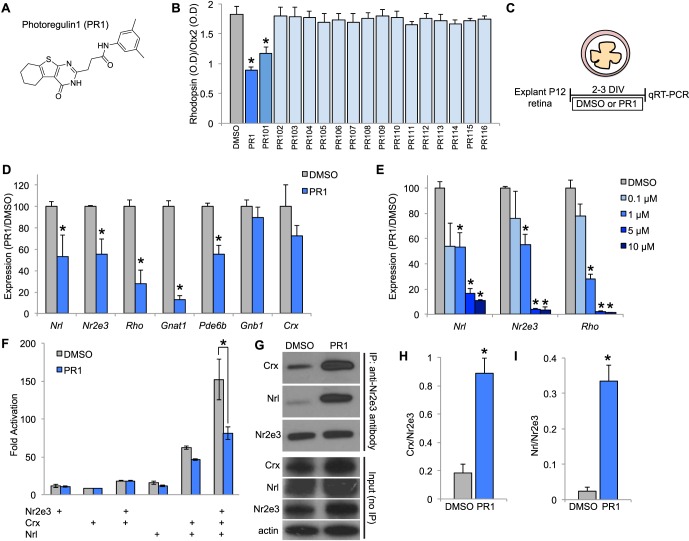
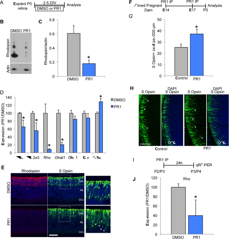
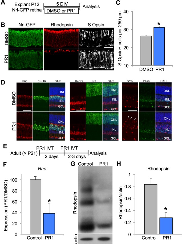
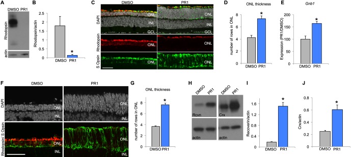
Purpose: Mutations in rod photoreceptor genes can cause retinitis pigmentosa (RP). Rod gene expression is regulated by the nuclear hormone receptor, Nr2e3. Genetic deletion of Nr2e3 reprograms rods into cells that resemble cone photoreceptors, and might therefore prevent their death from some forms of RP. There are no identified ligands for Nr2e3; however, reverse agonists might mimic the genetic rescue effect and may be therapeutically useful for the treatment of RP.

Methods: We screened for small molecule modulators of Nr2e3 using primary retinal cell cultures and characterized the most potent, which we have named photoregulin1 (PR1), in vitro and in vivo. We also tested the ability of PR1 to slow the progression of photoreceptor degeneration in two common mouse models of autosomal dominant RP, the RhoP23H and the Pde6brd1 mutations.

Results: In developing retina, PR1 causes a decrease in rod gene expression and an increase in S opsin+ cones. Photoregulin1 continues to inhibit rod gene expression in adult mice. When applied to two mouse models of RP, PR1 slows the degeneration of photoreceptors.

Conclusions: Chemical compounds identified as modulators of Nr2e3 activity may be useful for the treatment of RP through their effects on expression of disease-causing mutant genes.


Figures

Figure 1
Figure 1
(A) Chemical structure of PR1. (B) Effect of 1 μM PR1 or PR1 analogs PR101-PR116 on Rhodopsin expression (optical density) normalized to Otx2 expression (optical density) in dissociated retinal cell cultures (n = 4). *P < 0.05, ANOVA and Dunnett's tests. (C) Schematic showing experimental design for testing the effects of Nr2e3 modulators in intact retinal explant cultures for 2 to 3 DIV. (D) Reverse transcription qPCR for rod photoreceptor genes expressed in DMSO-treated controls and 1-μM PR1-treated retinal explants from P12 mice for 3 DIV (n = 4). *P < 0.05, Student's t-test. (E) Dose-response relationship of PR1 on rod-specific genes Nrl, Nr2e3, and Rhodopsin in P12 retinal explants for 2 to 3 DIV (n = 3 to 4). *P < 0.05 from DMSO treatment, Student's t-test. (F) HEK293T cells were transfected with Nr2e3, Crx, or Nrl and BR-225Luc (firefly luciferase driven by the bovine Rhodopsin promoter) and pRL-CMV (renilla luciferase driven by the CMV promoter; internal transfection control) and then treated with DMSO or PR1 10 μM for 2 days. We found PR1 decreased Rho promoter activity after transfection of Nr2e3, Crx, and Nrl (n ≥ 3). *P < 0.05, ANOVA, and Tukey's multiple comparison test. (G) Western blot analysis of DMSO and 10-μM PR1-treated HEK293T cells after transfection with Nr2e3, Crx, and Nrl followed by immunoprecipitation with an antibody generated against Nr2e3. Western blots for input (unprecipitated lysates) are shown below. (H) Quantification of Crx after immunoprecipitation with an anti-Nr2e3 antibody in DMSO and PR1-treated HEK293T cells (n = 3). *P < 0.05, Student's t-test. (I) Quantification of Nrl after immunoprecipitation with an anti-Nr2e3 antibody after treatment with DMSO or PR1 (n = 3). *P < 0.05, Student's t-test.
Figure 2
Figure 2
(A) Schematic showing experimental design for testing the effects of PR1 in intact retinal explant cultures from P0 mice (BE). (B) Western blot for rhodopsin shows significant reduction in P0 explants treated with PR1 1 μM for 5 DIV. (C). Rhodopsin expression was normalized to β-actin. Explants treated with PR1 had less relative expression than DMSO controls (n = 4). *P < 0.05, Student's t-test. (D) Reverse transcription qPCR for P0 explants treated with DMSO or PR1 1 μM for 2 DIV. We found Nrl, Nr2e3, Rhodopsin, and Gnat1 were significantly reduced with PR1, while Gnb1 and Crx did not change. The expression of the cone gene Thrb was increased in PR1-treated explants (n = 3). *P < 0.05, Student's t-test. (E) Sections from DMSO and 1-μM PR1-treated P0 explants stained for rhodopsin and S-opsin demonstrate a decrease in rhodopsin+ cells and an increase in S-opsin+ cells (arrows). Scale bar: 50 μm. F. Schematic for experimental design for (GH). Timed-pregnant dams were injected with PR1 at E14 and E17 and pups were euthanized for analysis at P0. (G) Pups treated with PR1 had more S-opsin+ cells per 200 μm of central retina compared with controls (n ≥ 6 retinas). *P < 0.05, Student's t-test. (H) Sections of central retina from control and PR1 pups stained for S-opsin (arrows) and DAPI. Scale bar: 50 μm. (I) Schematic for experimental design for (J). Postnatal pups were IP injected with DMSO or PR1 at P2 or P3 and then euthanized 24 hours later for RT-qPCR analysis. (J) Photoregulin1 decreased Rho expression in P2 and −3 pups (n ≥ 4). *P < 0.05, Student's t-test.
Figure 3
Figure 3
(A) Schematic for experimental design for (BD). Retinas from P12 Nrl-eGFP mice were explanted in DMSO or PR1 1 μM for 5 DIV. (B) Staining for rhodopsin and S-opsin, and GFP (unstained) in P12 explants from Nrl-GFP mice demonstrate a decrease in rhodopsin expression and an increase in S-opsin+ cells in PR1-treated retinas. Scale bar: 50 μm. (C) Photoregulin1-treated retinas had more S-opsin+ cells (arrows) than DMSO controls per 250 μm of central retina (n = 3). *P < 0.05, Student's t-test. (D) Quantification of bipolar cells (PKCα, Chx10); amacrine cells (HuCD); and Müller glia (Sox2) revealed no difference in the number of these cells (n = 3; P > 0.05, Student's t-test). However, we observed the migration of Sox2+ Müller glia into the ONL of PR1-treated retinas (arrows). (E) Schematic for experimental design for (FH). Adult mice received two intravitreal (IVT) injections 2 days apart of PR1 10 mM into one eye. (F) Compared with the control retina, PR1 decreased Rho expression in adult retinas (n = 3). *P < 0.05, Student's t-test. (G) Western blot for rhodopsin shows that IVT injection of PR1 decreases expression compared with the uninjected, contralateral retina of adult mice. (H) Rhodopsin expression was normalized to β-actin expression. Photoregulin1 decreased the relative expression of rhodopsin after IVT injection in adult mice (n = 5). *P < 0.05, Student's t-test.
Figure 4
Figure 4
(A) Retinas from P8 RhoP23H mice were explanted in DMSO or PR1 2 μM for 3 DIV. Western blot analysis shows that PR1-treated RhoP23H retinas have less Rhodopsin expression than DMSO controls. (B) Rhodopsin expression was normalized to β-actin expression. Photoregulin1–RhoP23H retinas had less relative expression of rhodopsin than DMSO-treated RhoP23H controls (n = 3). *P < 0.05, Student's t-test. (C) Retinas from P12-RhoP23H mice were explanted in DMSO or PR1 1 μM for 6 DIV. Staining with DAPI demonstrates that PR1-treated RhoP23H retinas had thicker ONLs in the central retina compared with DMSO-treated RhoP23H retinas. (D) Quantification of DAPI+ cells in the ONL of the central retina of DMSO and PR1-treated RhoP23H retinas (n = 5). *P < 0.05, Student's t-test. (E) Reverse transcription qPCR analysis of Gnb1 expression in DMSO and PR1-RhoP23H retinas suggests greater rod survival with PR1 treatment, since PR1 does not affect expression of this rod-specific transcript (n = 3). *P < 0.05, Student's t-test. (F) Retinas from P7 Pde6brd1 mice were explanted in DMSO or PR1 1 μM for 14 DIV. Retinas treated with PR1 had more DAPI+ cells in the ONL of the central retina than DMSO controls. (G) Quantification of DAPI+ cells in the ONL of DMSO and PR1-treated Pde6brd1 retinas (n = 4). *P < 0.05, Student's t-test. (H) Retinas from P7 Pde6brd1 mice were explanted in DMSO or PR1 1 μM for 12 DIV and analyzed by Western Blot. Photoregulin1-treated retinas expressed more recoverin and Crx than DMSO controls, indicating greater photoreceptor survival. (I) Recoverin expression was normalized to β-actin expression. Photoregulin1-Pde6brd1 retinas expressed more relative recoverin than DMSO-Pde6brd1 retinas (n = 3). *P < 0.05, Student's t-test. (J) Expression of Crx was normalized to β-actin expression. Photoregulin1-treated retinas had greater relative expression of Crx than DMSO controls, indicating greater photoreceptor survival (n = 3). *P < 0.05, Student's t-test.

Similar articles

Cited by

References

    1. Hartong DT,, Berson EL,, Dryja TP. Retinitis pigmentosa. Lancet. 2006; 368: 1795– 1809. - PubMed
    1. Nguyen AT,, Campbell M,, Kiang AS,, Humphries MM,, Humphries P. Current therapeutic strategies for P23H RHO-linked RP. Adv Exp Med Biol. 2014; 801: 471– 476. - PubMed
    1. Gorbatyuk MS,, Knox T,, LaVail MM,, et al. Restoration of visual function in P23H rhodopsin transgenic rats by gene delivery of BiP/Grp78. Proc Natl Acad Sci U S A. 2010; 107: 5961– 5966. - PMC - PubMed
    1. Mao H,, Gorbatyuk MS,, Rossmiller B,, Hauswirth WW,, Lewin AS. Long-term rescue of retinal structure and function by rhodopsin RNA replacement with a single adeno-associated viral vector in P23H RHO transgenic mice. Hum Gene Ther. 2012; 23: 356– 366. - PMC - PubMed
    1. Carter-Dawson LD,, LaVail MM. Rods and cones in the mouse retina. I. Structural analysis using light and electron microscopy. J Comp Neurol. 1979; 188 245– 262. - PubMed

Publication types

MeSH terms