. 2017 Jan 31;8(5):7521-7532.
doi: 10.18632/oncotarget.13485.

Molecular mechanism and therapeutic implications of selinexor (KPT-330) in liposarcoma

Affiliations

Molecular mechanism and therapeutic implications of selinexor (KPT-330) in liposarcoma

Manoj Garg et al. Oncotarget. .

Abstract

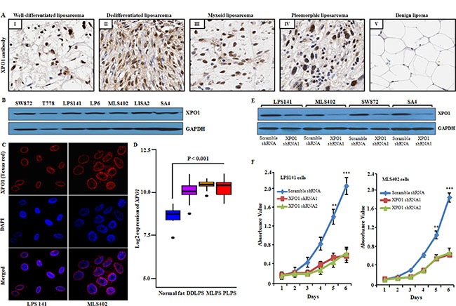
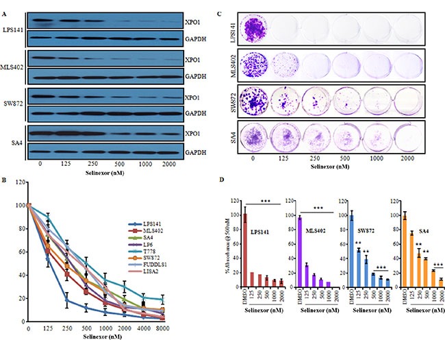
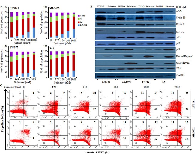
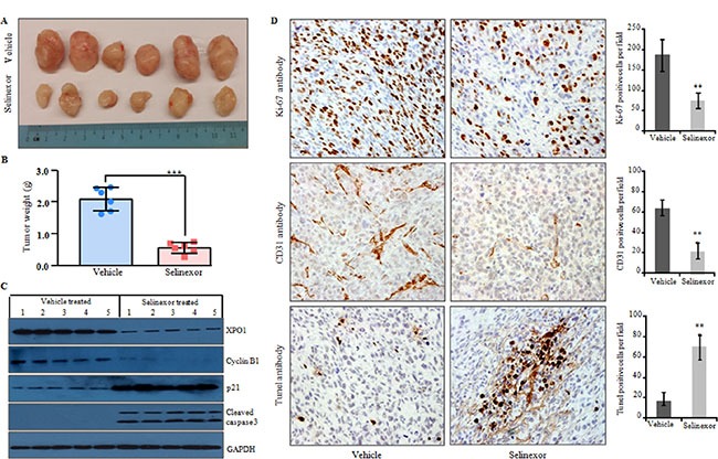
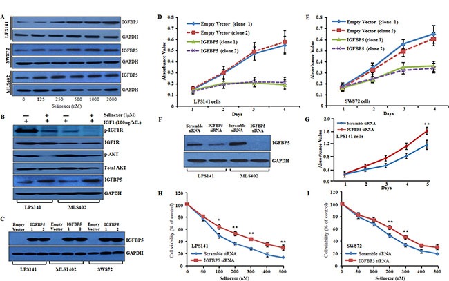
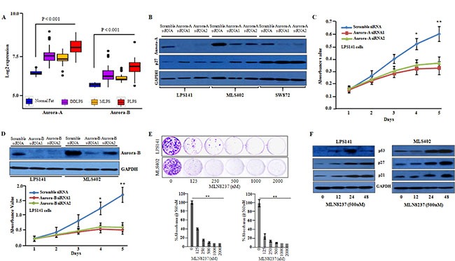
Exportin-1 mediates nuclear export of multiple tumor suppressor and growth regulatory proteins. Aberrant expression of exportin-1 is noted in human malignancies, resulting in cytoplasmic mislocalization of its target proteins. We investigated the efficacy of selinexor against liposarcoma cells both in vitro and in vivo. Exportin-1 was highly expressed in liposarcoma samples and cell lines as determined by immunohistochemistry, western blot, and immunofluorescence assay. Knockdown of endogenous exportin-1 inhibited proliferation of liposarcoma cells. Selinexor also significantly decreased cell proliferation as well as induced cell cycle arrest and apoptosis of liposarcoma cells. The drug also significantly decreased tumor volumes and weights of liposarcoma xenografts. Importantly, selinexor inhibited insulin-like growth factor 1 (IGF1) activation of IGF-1R/AKT pathway through upregulation of insulin-like growth factor binding protein 5 (IGFBP5). Further, overexpression and knockdown experiments showed that IGFBP5 acts as a tumor suppressor and its expression was restored upon selinexor treatment of liposarcoma cells. Selinexor decreased aurora kinase A and B levels in these cells and inhibitors of these kinases suppressed the growth of the liposarcoma cells. Overall, our study showed that selinexor treatment restored tumor suppressive function of IGFBP5 and inhibited aurora kinase A and B in liposarcoma cells supporting the usefulness of selinexor as a potential therapeutic strategy for the treatment of this cancer.

Keywords: IGFBP5; cell cycle; selinexor; xenograft.


Conflict of interest statement

CONFLICTS OF INTEREST

S.S, E.B, and M.K are employees and stockholders of Karyopharm Therapeutics. The remaining authors have no competitive financial interest.

Figures

Figure 1
Figure 1. Expression of XPO1 in human liposarcoma tissue and cell lines, and XPO1 knockdown in liposarcoma cells
(A) XPO1 protein expression was examined in liposarcoma tissue and benign lipoma using immunohistochemical analysis. Representative photomicrographs showed nuclear expression of endogenous XPO1 in well-differentiated liposarcoma (I), dedifferentiated liposarcoma (II), myxoid liposarcoma (III) and pleomorphic liposarcoma (IV) tissue samples, whereas benign lipoma (V) showed either very less or no reactivity (original magnification, X200; objective, X20). (B) Western blot analysis of liposarcoma cell lines probed with a XPO1 antibody (band 123 kDa, corresponding to the size of XPO1 protein). GAPDH used as the loading control. (C) Nuclear localization of XPO1 protein (red color) in fixed/permeabilized liposarcoma cell lines. DAPI (blue color) was used to stain nuclei. (D) Microarray data (GSE21122) from GEO database for samples of 46 dedifferentiated liposarcoma (DDLPS), 20 myxoid liposarcoma (MLPS), 23 pleomorphic liposarcoma (PLPL) and 9 normal fat tissue; approximately 90% of samples showed significant (P < 0.001) upregulation of XPO1 compared to normal fat samples. (E) Western blot confirmed knockdown of XPO1 protein in LPS141, MLS402, SW872 and SA4 cells infected with XPO1 shRNA1 compared to scrambled shRNA. GAPDH antibody was used to assure equal loading of lysates. (F) XPO1knockdown suppressed cell growth of LPS141, MLS402. Data represent mean ± SD; n = 4. **P ≤ 0.001, ***P ≤ 0.0001.
Figure 2
Figure 2. Selinexor significantly suppressed growth of liposarcoma cell lines in liquid culture
(A) LPS141, MLS402, SW872 and SA4 cells were treated with either diluent (DMSO) or increasing concentrations of selinexor (0, 125, 250, 500, 1000 and 2000 nM, 24 h). Cell lysates were analyzed by western blots for XPO1 protein (GAPDH, internal control). (B) Selinexor inhibited cell proliferation of liposarcoma cell lines in a dose-dependent manner. Cells were cultured in the presence of selinexor at the indicated doses for 72 hours, and cell growth was assessed by MTT assay. Data represent mean ± SD; n = 4. (C and D) Selinexor suppressed clonogenic growth. Cells were treated with indicated concentration of selinexor for 24 h, washed and then allowed to form colonies for 14 days. Colonies were stained with crystal violet and dissolved in DMSO. Representative photomicrograph (C) and quantitative analysis showed a reduction in clonogenic growth (D). Data are expressed as mean values ± SD of at least four independent experiments. **P ≤ 0.001, ***P ≤ 0.0001.
Figure 3
Figure 3. Selinexor induced cell cycle arrest and apoptosis in liposarcoma cell lines
(A) Liposarcoma cells were incubated with either diluent control (DMSO) or different concentrations of selinexor for 48 h, stained with propidium iodide (PI) to determine cell cycle profiles using flow cytometric analysis. Data displayed as histogram (mean of three independent experiments). (B) Liposarcoma cells were cultured in presence of selinexor (1000 nM) for 24 h. Cell lysates were prepared and subjected to western blot analysis using different antibodies (GAPDH, loading control). (C) Apoptosis of liposarcoma cells after 48 h exposure to different concentration of selinexor. Cells were stained with Annexin V-FITC and PI and analyzed by flow cytometry. Percentage of apoptotic cells either Annexin V + PI or both is displayed in each treatment group of three independent experiments.
Figure 4
Figure 4. Selinexor significantly reduced tumor growth of LPS141 cells in a xenograft murine model
LPS141 cells (2 × 10⁶) were implanted subcutaneously into the right flank of 6-week old male NSG mice. After 14 days, mice were randomly placed into two groups and treated by gavage with either vehicle control or selinexor (10 mg/kg, twice a week X 4 weeks). (A) Tumors from mice treated with vehicle versus selinexor (n = 6 for each group). Scale in cm. (B) Selinexor significantly reduced tumor weight compared to vehicle (dissected tumors). Data are the mean ± SD of the tumors. ***P ≤ 0.0001 (Student's t-test). (C) Western blot analysis of lysates of LPS141 xenograft tumors from mice treated with selinexor; protein expression of XPO1, cyclin B1, p21, cleaved caspase 3 (GAPDH, internal control). (D) Immunohistochemical staining of Ki-67 (proliferation), CD31 (blood vessels) and TUNEL (apoptosis) in liposarcoma xenograft tumors from mice treated with either vehicle or selinexor (original magnification, X 200; objective, X 20. Columns (on the right) show percentage positively stained cells (mean ± SD of three independent experiments). **P ≤ 0.001(Student's t-test).
Figure 5
Figure 5. Inhibition of XPO1 induced cytotoxicity by re-expressing IGFBP5; and IGFBP5 overexpression reduces cellular proliferation, migration and invasion in liposarcoma cell lines
(A) IGFBP5 protein expression by western blot analysis in LPS141, SW872 and MLS402 following selinexor treatment (0–2000 nM, 24 h) (GAPDH, internal control). (B) LPS141 and MLS402 cells were serum-starved overnight, treated with selinexor (1000 nM) for 2 h and then stimulated with human IGF-1 (100 ng/ml) for 10 minutes, and the proteins were analyzed by western blot using indicated antibodies. (C) Western blot shows overexpression of IGFBP5 protein in LPS141, MLS402 and SW872 cells stably transfected with IGFBP5expression vector compared to empty vector (GAPDH, loading control). Clone 1 and 2 are two separate clones that stably express IGFBP5. (D and E) Overexpression of IGFBP5 in LPS141 and SW872 cells exhibited decreased cell growth in liquid culture. Clones 1 and 2 were two different stable clones expressing IGFBP5. For control, two separate clones containing empty vector were used. Data represent mean ± SD; n = 4. **P ≤ 0.001; ***P ≤ 0.0001 (Student t test). (F) Western blot analysis verified silencing of IGFBP5. (G) MTT assay showed that knockdown of IGFBP5 increased cell proliferation in liquid culture. (H and I) LPS141 and SW872 cells were transfected either with IGFBP5 siRNA or scramble siRNA. These cells were exposed to different concentration of selinexor for 48h, and growth inhibition was measured by MTT assay. Data represent mean ± SD; n = 4. **P ≤ 0.001. Data for G, H and I represent mean ± SD of three independent experiments done in triplicates. **P < 0.001 (Student's t-test).
Figure 6
Figure 6. Inhibition of aurora-A and aurora-B decreased the cellular growth of liposarcoma cells
(A) Microarray data (GSE21122) showed that aurora-A and aurora-B were significantly upregulated in liposarcoma patient samples compared to normal fat. (B) Western blot analysis confirmed knockdown of aurora-A in siRNA1 and siRNA2 transfected cells compared to scramble siRNA in LPS141, MLS402 and SW872 cells. (C) Aurora-A siRNAs suppressed the growth of LPS141 cells in liquid culture. Data represent mean ± SD; n = 4. *P ≤ 0.01; ***P ≤ 0.001 (Student t test). (D) Western blot analysis confirmed silencing of aurora-B in LPS141 and MLS402 cells and aurora-B knockdown suppressed the growth of LPS141 cells in liquid culture. (E) Soft agar assay: MLN8237 significantly inhibited clonogenic growth of LPS141 and MLS402. Data represent mean ± SD of three independent experiments done in triplicates. **P < 0.001 (Student's t-test). (F) LPS141 and MLS402 cells were treated with MLN8237 (500 nM) for 12, 24, 48 h; and the protein levels of p53, p21, p27 were analyzed by western blot. GAPDH was used to ensure equal loading of lysate.

Similar articles

Cited by

References

    1. Crago AM, Singer S. Clinical and molecular approaches to well differentiated and dedifferentiated liposarcoma. Curr Opin Oncol. 2011;23:373–8. - PMC - PubMed
    1. Dalal KM, Kattan MW, Antonescu CR, Brennan MF, Singer S. Subtype specific prognostic nomogram for patients with primary liposarcoma of the retroperitoneum, extremity, or trunk. Ann Surg. 2006;244:381–91. - PMC - PubMed
    1. Kanojia D, Nagata Y, Garg M, Lee DH, Sato A, Yoshida K, Sato Y, Sanada M, Mayakonda A, Bartenhagen C, Klein HU, Doan NB, Said JW, et al. Genomic landscape of liposarcoma. Oncotarget. 2015;6:42429–44. doi: 10.18632/oncotarget.6464. - DOI - PMC - PubMed
    1. Gutierrez A, Snyder EL, Marino-Enriquez A, Zhang YX, Sioletic S, Kozakewich E, Grebliunaite R, Ou WB, Sicinska E, Raut CP, Demetri GD, Perez-Atayde AR, Wagner AJ, et al. Aberrant AKT activation drives well-differentiated liposarcoma. Proc Natl Acad Sci USA. 2011;108:16386–91. - PMC - PubMed
    1. Kransdorf MJ. Malignant soft-tissue tumors in a large referral population: distribution of diagnoses by age, sex, and location. AJR Am J Roentgenol. 1995;164:129–34. - PubMed

MeSH terms